What is a petri dish used for? Petri dish and its history. Preparation of petri dishes
pure cultures
In 1872, Robert Koch, the future great microbiologist, was appointed health officer in Wolstein (now Wolsztyn in Poland), where anthrax was rampant at that time. This disease has been known since antiquity under the name "sacred fire": people believed that only angry gods could send such punishment to the earth. Anthrax was a threat to all agriculture - livestock most often fell ill. However, not only animals died, but also people: farmers, shepherds, milkmaids.
Examining the blood of dead animals under a microscope, Koch discovered that only one microbe, the bacillus, was to blame for the development of the disease ( Bacillus anthracis). The scientist was able to isolate the bacillus and grow pure culture- a culture represented by one type of microbes. He infected a perfectly healthy animal with a pure culture, giving it anthrax. The scientist realized that the creation of pure cultures is the key to successful determination of the causes of infection.
Robert Koch, like his predecessors, grew bacteria in a liquid medium - meat or grain broths. Koch managed to obtain a pure culture of anthrax bacillus in liquid broth, but he was looking for another method. There were good reasons for this. If several types of bacteria got into the broth, they mixed with each other and it was extremely difficult to separate them. I had to transplant bacteria more than once. From the solution, where the necessary bacteria were the most, Koch took a small drop and transferred it to a fresh broth. There were already fewer foreign bacteria in the new broth, but this operation had to be monotonously repeated several times, so that as a result only one type of microbes appeared in the nutrient medium.
Robert Koch(1843–1910) German microbiologist. He discovered anthrax bacillus, vibrio cholerae and tuberculosis bacillus (Koch's bacillus). In 1905 he was awarded the Nobel Prize in Physiology or Medicine "for research and discoveries concerning the treatment of tuberculosis."

Before Robert Koch, researchers observed microorganisms as colorless, which led to numerous errors. Koch used aniline dyes that selectively stain only microbes. After Koch's experiments, scientists around the world began to invent methods for staining bacteria. So, in 1884, the doctor Hans Christian Gram came up with a staining method that became one of the main methods for determining the presence and type of bacteria in a particular substrate.
Why are pure cultures needed?
Microbes are very small (on average 0.5-5 microns), do not differ much in appearance, which causes certain difficulties in their study. For research, it is necessary to isolate a microbe from the surrounding world, full of a variety of microorganisms. A microbial cell, once on a nutrient medium, gives offspring - a clot of identical cells, a colony, which can be studied as a single microorganism. It turned out that, by choosing the cultivation conditions, it is possible to obtain pure cells of any microorganism. And that means giving it a name, describing its properties, classifying it. Thanks to this discovery by Robert Koch, microbiology was singled out as an independent science.
solid medium
After the report on the discovery of the causative agent of anthrax, Robert Koch was invited to head the laboratory at the microbiological institute in Berlin and was offered the position of adviser to the Ministry of Health. Koch had good equipment, talented assistants and the opportunity to solve a riddle that had tormented him for a long time. It was known that tuberculosis also causes some kind of microbe: it was possible to infect healthy animals with the tissues of a sick person. Koch managed to pick up a technique for staining tissue preparations in order to see the pathogen bacteria under a microscope. But his joy was short-lived - the bacterium did not want to grow on ordinary nutrient media.

Macro shot of tubercle bacillus colonies ( Mycobacterium tuberculosis). They are distinguished by a colorless uneven surface.
Once a scientist noticed a moldy potato thrown on the table with an abundance of multi-colored specks-colonies - gray, yellow, green. He collected samples from each colony and saw under the microscope that each speck is a colony of one type of microbe! In a liquid medium, the microbes mixed and it was extremely difficult to separate them. And on a solid medium, they remained in one place, multiplied and gave a pure culture!

Koch's accidental observation made a revolution: fresh potatoes became one of the first solid media for growing microorganisms. However, such a nutrient substrate is not suitable for all microbes, so the search for an alternative solid medium continued.
Koch again diligently began to grow tuberculosis culture. The bacterium did not grow on potato slices. Then he began to use gelatin to turn the broth into a solid nutrient medium. After many unsuccessful attempts, Koch added blood serum to the medium in order to recreate the conditions of a living organism. After 15 days (an unusually long time for the anthrax bacillus), droplets of colonies of the dangerous tubercle bacillus appeared on the surface of the medium.
Microorganisms divide every 20 minutes, so already 3 hours after transplanting microbes on a Petri dish, you can see colonies, and a day later the number of bacteria in them is in the millions.
microbiological art
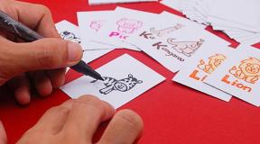
Alexander Fleming came up with a new art form - drawing bacteria on a solid medium. Fleming, as a member of the Chelsea Art Club, invented amateur watercolors. The scientist painted graceful ballerinas, luxurious houses, soldiers with bacteria. The creation of the picture required accuracy: it was necessary not only to find bacteria with different pigments, but also to choose the growing time so that the same colors would grow at the same time and not violate the color boundaries.

The American Society for Microbiology (ASM), inspired by the example of Fleming and his colleagues, has been holding the Agar Art Competition since 2015. Talented microbiologists and artists paint real pictures. Some come up with their own stories, others reproduce the paintings of the masters, for example, Van Gogh's Starry Night.

In 1928, the British bacteriologist Alexander Fleming discovered that a colony of mold fungi had grown next to bacteria on agar in one of the Petri dishes. The bacterial colonies around the mold turned pale - their cells were destroyed. Fleming isolated from mold fungi a substance that destroys bacterial cells - penicillin, the first antibiotic. Fleming's discovery changed not only medical science, but also the fate of many seemingly hopeless patients.
Talented laboratory assistant
At that time, the only laboratory bottle that was suitable for growing microbes was a test tube. But handling it required skill: if you put the test tube horizontally, the unsolidified medium will spill, if you put it at an angle, it can fall and break. The chance of contracting a dangerous disease multiplied before our eyes! Then Koch and his laboratory assistants came up with the idea of pouring the nutrient medium into cups and covering them with tall glass caps. But to look at the colonies, the cap had to be removed, and this is a direct route of infection. It was then that the finest hour of one of the laboratory assistants came - Julius Petri. He reduced the height of the walls of the cup in which the microbes were grown, leaving low sides. And instead of a huge cap, he covered it with another transparent cup - it turned out to be more convenient to observe the colonies.
Petrie worked under Koch for only a couple of years (1877–1879), but during that time he had a profound effect on the future of microbiology. The innovation of Julius Petri gave a strong impetus to medicine and saved millions of lives. After working under Robert Koch, he headed the Gobersdorf sanatorium, the first European center for the treatment of tuberculosis patients.
agar agar
In Koch's laboratory, not only a Petri dish appeared, but also a standard filling for it - an agar nutrient medium. Its predecessor, a medium containing gelatin, melted easily when heated, like jelly put in a warm place. Colonies on such a medium turned into mush.
Agar-based medium invented Walter Hesse, another laboratory assistant of Koch. He entered the service along with his wife Fanny. She was not listed as an employee of the laboratory, but she did the work of a scientific illustrator - she sketched the microorganisms that she saw under a microscope.
One day Fanny made jelly. Hesse noticed that it does not melt in the sun and retains its shape. He found out that the main component of jelly is agar-agar, a substance that is extracted from red and brown algae. Walter replaced the standard gelatin with it, and the bacterial culture media became harder. Agar-agar is still used today for the preparation of media, but it is purified in a special way.

To determine the effectiveness of antibiotics in the pharmaceutical industry, special tests are used. For example, the agar diffusion method. Microorganisms are inoculated into Petri dishes. Disks containing certain doses of different antibiotics are placed on the seeded surface at an equal distance from each other. The larger the radius of the growth inhibition zone, the more effective the drug against this microorganism.
At first, the Petri dish was used only for growing cell cultures, but now these dishes are used in various fields of science. For example, Petri dishes are used to study the effectiveness of antibiotics, they are used to study food safety and grow genetically engineered bacteria that synthesize the insulin necessary for diabetics.
Petri dishes are made in different sizes and from a variety of materials - glass, plastic and even stainless steel. For work, the scientist can choose the right one.
Do you know how many bacteria live on your palms? And on the cover of the phone? And on some item that you use every day and, as you think, keep clean? Not? Would you like to see? We offer an affordable way to see the world that lives at your fingertips.
What you need
1. Broth:
- fatty meat (50 g);
- water (0.5 l);
- a bag of agar (this substance is isolated from red and brown algae, sold in some grocery stores in powder form).
2. Sterile Petri dishes (5-10 pcs.):
This is such a glass or plastic laboratory glassware, consisting of two flat cups that fit into each other. This structure serves to protect against external pollution or gases released by the bacterial colonies themselves. Petri dishes are sold in most pharmacies, they are cheap.
3. Refrigerator.
4. Thermometer.
5. Cotton swabs (20 pcs.).
6. Antibacterial soap (for testing).
7. Mask, gloves.
8. Chlorine solution, for example, "Whiteness" liquid (0.5 l).
Sequencing
Building a house
1. Prepare an environment for the growth and reproduction of future bacteria: boil the meat broth. It is better to cook the meat longer to get richer.
2. Add 1/2 teaspoon (1.2 g) of agar to the boiled broth for every 1/4 cup (60 ml of liquid). This amount is suitable for one Petri dish.
3. Microwave your stock, bring to a boil, and wait until all the agar powder has dissolved. Cool down afterwards.
4. Take Petri dishes. Please note: they must be sterile inside! If you are not sure, dip in boiling water, but be careful not to scald yourself. In order not to bring bacteria from the air, it is better to do all operations near a burning gas burner.

5. Pour the nutrient mixture into the bottom half in a thin layer, just lightly covering the bottom, and cover the container with the top cup as quickly as possible. Let cool.

6. Place the closed Petri dishes in the refrigerator. It is better that the bottom half is on top. This is necessary to prevent the destruction of the medium by drops of condensate. Wait until the medium becomes like a hard jelly. Finished Petri dishes can be stored in the refrigerator for about two months.
housewarming
1. Remove the Petri dishes from the refrigerator and warm them to room temperature. Now you can start moving in! There are many options - it all depends on your imagination. We decided to see what bacteria live on our fingertips.

2. Touch the jelly alternately with five fingers in different places. This must be done carefully, without damaging the surface. Cover the container with fingerprints with the top cup (this must be done very quickly) and seal, for example, with adhesive tape. On the underside of the cup, circle the fingerprints with a marker and sign.

3. Put the inhabited "houses" in a warm and dark place. With the second point, everything seems to be clear, but as for heat, bacteria are quite picky and grow most actively at +37 degrees. In order to determine the optimal mode, take a thermometer and measure the temperature of warm surfaces in the house - for example, behind the refrigerator. The growth time in this case will be 1-2 days. It is possible to grow bacteria in cooler conditions, but then the process will be much slower.

4. Fix the results: after a day, with a marker, start drawing circles on the outside of the bottom of the cup around the overgrown bacteria, mold, fungi or other "tenants". So you will receive daily information on how and at what speed the “living space” is populated (perhaps a drawing will appear that resembles a cut of a tree).

5. Try to identify the microorganisms that have grown in colonies on the plates. To do this, microbiologists have many ways such as isolating a pure culture, Gram stain, biochemical samples. We will be able to use only a few - to determine the color and shape of the colony.

6. Check the effectiveness of antibacterial soap. As soon as life develops in the petri dish, apply a drop of soap between the two colonial islands with a Q-tip. Close the cup and leave it for another day in a warm and dark place. The areas of settlement will grow around the drop at some distance from it, and the more active the substance, the wider the “exclusion zone” will be.
Acquaintance
You can simply leave your fingerprint (then you only need one Petri dish). Or you can conduct an experiment and compare the composition of the “residents” in different situations (for this you will have to use several cups), for example:
- after a trip in public transport;
- after long work at the computer;
- after hands are washed with / without soap;
- after returning from work / school / university;
- after shaking hands with neighbors on the right, left and below, as well as riding in an elevator with the owner of three American bulldogs...
For comparison, leave prints of the cleanest hands in one of the cups (wash with antibacterial soap, wipe with a disinfectant solution).
There are many different types of microorganisms living on your fingers, and it is very difficult to determine what has grown in which place. We can only make assumptions, for example:
- different types of staphylococci can form dyes (pigments) of golden, white and lemon yellow colors;
- colonies of Serratia marcescens are red;
- pink micrococcus - pink;
- Pseudomonas aeruginosa - blue;
- chromobacter violet - violet;
- mycobacterium tuberculosis - yellow or bright orange;
- some types of mold (just in case, we remind you that mold is a type of fungus, not bacteria) form black hairy colonies.
Of course, microbes can only be accurately identified in the laboratory.
Eviction
Sooner or later, the colony will fully satisfy your curiosity and, according to the law of microorganism growth, will begin to die. And this means that the time has come to get rid of it - harshly and mercilessly, in order to free the house for new tenants. Well, or decide that being a microbiologist is not your calling, and get rid of Petri dishes. In fact, colonies that have grown at the wave of your hand are hardly dangerous, but this does not relieve you of the obligation to bury them according to all the rules of microbiology with all rituals and honors.

1. Protect yourself by wearing an apron, mask and rubber gloves.

2. Fill Petri dishes with a small amount of chlorine solution and leave for a few minutes.

3. Pour out the contents, rinse the cups, put them in bags, and then in the trash can.
A warning: If you want to continue experimenting with microbial growth yourself, do not use any biological fluids (such as saliva), otherwise you can grow colonies that are dangerous to health. Take precautions and be careful!
Modifications
The Petri dish is usually made of clear glass or plastic (clear polystyrene) and comes in a variety of sizes. The most commonly used options have a diameter of about 50-100 mm and a height of about 15 mm.
Application
The cup is widely used in microbiology for culturing colonies of microorganisms. To do this, it is filled with a layer of nutrient medium, on which the culture of microorganisms is sown.
Glass dishes are reusable but require sterilization before re-inoculation. Cups made of plastic materials are supplied sterile, in sealed packaging.
In addition, the Petri dish is often used for applied purposes, for example, for evaporating liquids, storing small fragments of various preparations, dissecting small animals and plants, and etching small printed circuit boards.
Often, Petri dishes are used in terrariums for a “nesting base” when breeding amphibians (dart frogs, leaf climbers and other animals).
see also
|
||||||||||||||||||||||
Write a review on the article "Petri Dish"
An excerpt characterizing the Petri dish
- I told you - this cave is evil! .. I told ... I told you! - convulsively sobbing, the kid lamented - Well, why didn’t you listen to me! And what should I do now?.. Where should I go now?..Tears flowed down dirty cheeks in a burning stream, tearing apart a small heart... Beloyar didn't know if his beloved grandfather was still alive... Didn't know if the evil people would come back? He was just scared as hell. And there was no one to comfort him... no one to protect him...
And Svetodar lay motionless at the very bottom of a deep crack. His wide-open, clear blue eyes, seeing nothing, looked at the sky. He went far, far away, where Magdalena was waiting for him... and his beloved father with kind Radan... and sister Vesta... and his gentle, affectionate Margarita with her daughter Maria... and unfamiliar granddaughter Tara... And all- all those who died long ago defending their native and beloved world from non-humans who called themselves humans...
And here, on the ground, in a lonely empty cave, on a round pebble, hunched over, a man was sitting... He looked quite small. And very scared. Bitterly, weeping angrily, he furiously rubbed his evil tears with his fists and swore in his childish soul that such a day would come when he would grow up, and then he would certainly correct the “wrong” world of adults ... Make it joyful and good! This little man was Beloyar... a great descendant of Radomir and Magdalena. Small, lost in the world of big people, crying Man...
Everything I heard from the lips of the North once again flooded my heart with sadness ... I asked myself again and again - are all these irreparable losses really natural? .. Is there really no way to rid the world of evil spirits and malice ?!. All this terrible machine of global killing made the blood run cold, leaving no hope of salvation. But at the same time, a powerful stream of life-giving force flowed from somewhere into my wounded soul, opening every cell in it, every breath to fight against traitors, cowards and scoundrels! .. With those who killed the pure and brave, without hesitation, by any means, if only to destroy everyone who could be dangerous for them ...
Petri dish. Anyone who has ever dealt with medical or biological research knows what it is. A round and flat glass plate with a lid, 5 or 10 cm in diameter, designed for growing laboratory microorganisms was invented in 1877 by the German bacteriologist Julius Richard Petri, assistant to Robert Koch. Now it literally grows works of art. Scientific Russia talks about the joint work of scientists and bacteria.
For over 100 years, it has been the most familiar laboratory glassware. A heated liquid nutrient medium is poured into it - agar-agar - and it solidifies into a flat, translucent grayish plate. On this nutrient medium, scientists “sow” microorganisms. Microbes like to live in Petri dishes, they have everything they need to live there - food and the warmth of a thermostat. Colonies of bacteria grow, taking on a variety of shapes and colors. You can “sow” microbes simply by opening a Petri dish with agar, for example, in a student canteen. And everything that gets into it with air will bloom in a lush color in a matter of days. This experience is often practiced by undergraduate students.
However, new times have made the Petri dish a real art object. Who and when first came up with the idea to make the bizarre forms of bacterial colonies an object of art - history is silent. Perhaps the first was the famous Alexander Fleming, a British microbiologist who discovered the antibacterial enzyme lysozyme produced by the human body and was the first to isolate penicillin from the fungus Penicillium notatum - historically the first antibiotic.
Fleming was famous among his colleagues for the creative mess in the laboratory and luck. Once, when the doctor had a cold, he sowed the mucus from his own nose onto a Petri dish that already contained bacteria, and after a few days found that the bacteria were destroyed in the places where the mucus had been applied. This is how lysozyme was discovered, and the first article about it was published in 1922. And in 1928, he discovered that a colony of molds had grown in one of the Petri dishes containing Staphylococcus aureus bacteria. Colonies of bacteria around the molds became transparent due to cell destruction. This is how penicillin was discovered, which saved millions of lives. As a true British scientist, Fleming was no stranger to originality, and on one of his Petri dishes he left such a masterpiece.

Nowadays, there are already several recognized masters of agar art - a new type of creative self-expression of scientists, and not only biologists. In 2015, the American community of microbiologists organized the first international competition "The Agar art" (Agar art). The winning entry was "Neurons" by British microbiologist Maria Penil of New England Biolabs. After growing the bacteria for two days at 30°C, the author usually leaves them alone for a few more days. After that, the result is fixed with epoxy resin.
In second place is the bacterial map of New York, created at New York City's Community Biolab. There are no longer classic round Petri dishes, but square glass plates with bacterial cultures taken from more than 50 New Yorkers.

Rounding out the top three is Harvest Season by Argentinean microbiologist Maria Eugenia Inda of Cold Spring Harbor Labs. The work uses yeast bacteria, which are the basis of such products as bread, wine, beer, and so on. The picture shows a modest farmhouse with a yeast field surrounding it. "This yeast field is ripe for harvest!" - comments the author of the work.
A true master of agar art, who has found his own way of growing unique masterpieces, is the Israeli scientist Eshel Ben-Jakob, a professor of physics at Tel Aviv University in Israel.


He found a way to grow the shoots of microbial colonies in a targeted manner, by regulating the areas where nutrients are found. In his works, the colonies themselves "stretch" with a kind of "antennae" or "tentacles" to the source of nutrients. The scientist-artist paints the resulting amazing forms in different colors.

Eshel Ben-Jakob is one of the authors of the work on biocommunication in microorganisms. The scientist believes that bacteria have a special kind of collective behavior and a primitive form of social consciousness. The experiment shows that when a colony enters extreme conditions of nutrient deficiency, exposure to antibiotics, or exit from the optimum temperature, microorganisms themselves reduce their own population, releasing a special substance that kills part of the colony. Bacteria communicate, according to Ben-Jakob, using a "chemical language" that allows them to turn colonies into a large "brain". It is he who helps bacteria respond so effectively to environmental changes.

Hunter Cole of Chicago's Loyola University (USA) creates compositions from several Petri dishes inoculated with bioluminescent bacteria. She photographs them at different stages of the life cycle of the colonies, and amazing luminous compositions are obtained.

Scientists go even further in creating amazing new art that is part of the research process. They use genetic engineering methods to achieve the emergence of new bacterial pigments and fluorescent proteins, directed changes in the structure of bacterial colonies. At some point, you begin to understand that science and art are two absolutely inseparable parts of one amazing creative process that complement each other and create a unique result.