What the human cardiovascular system consists of. The structure and function of the organs of the cardiovascular system. What is the cardiovascular system
Briefly, the following can be distinguished: the heart and the network of blood vessels.
The cardiovascular system is basic in human anatomy. In addition to the blood supply to all organs, it performs a regulatory function, and also unites the subsystems of our body into a single whole.
The structure and function of the human heart
In short, the human cardiovascular system has the typical characteristics of mammals. First, the human heart consists of four special chambers with symmetry - the right and left atrium, right and left ventricle. Different vessels enter different atria: in the left - pulmonary veins, in the right - hollow. Different arteries also leave the ventricles: from the left - the ascending aorta, from the right - the pulmonary artery.
As a hollow muscular organ, the heart has layers that are different in structure and purpose. The epicardium, or the outer lining of the heart, protects it from infections. The myocardium provides quality contractions. The endocardium lines the inner surface, due to its folds, heart valves are formed, which form the correct blood flow.
In order for the heart to work harmoniously, it has a conducting system. It is formed from special muscle fibers, as well as nodes and bundles, consisting of fibers. In their structure, the fibers resemble a combination of muscle and nervous tissue. Due to the coordination of contractions of the parts of the heart, the conducting system ensures the automatism of the heart and the rhythm of its contractions.
Blood vessels: what are they for?
The structure of the vascular system is extremely complex. The vessels provide the movement of blood, pushed out by the heart, in two circles of blood circulation. The first - large - begins in the left ventricle and ends in the right atrium. The wall of the left ventricle is three times thicker than the right one. This is due to the fact that the task of the systemic circulation is the blood supply to all organs. Therefore, the left ventricle needs to make significant efforts to ensure the expulsion and subsequent movement of blood along a long path. The time for the blood to pass through the great circle is less than half a minute. The second circle of blood circulation is called small, and ensures the movement of blood only in the vessels that wash the lungs. Due to the small circle of blood circulation, the blood is saturated with oxygen. It starts in the right ventricle and ends in the left. In a small circle, blood moves much faster than in a large circle - the circulation time is only 4-5 seconds.
Large and small circle of blood circulation
- Characteristics of the cardiovascular system
- Heart: anatomical and physiological structural features
- Cardiovascular system: vessels
- Physiology of the cardiovascular system: systemic circulation
- Physiology of the cardiovascular system: scheme of the pulmonary circulation
The cardiovascular system is a collection of organs that are responsible for ensuring the circulation of blood flow in the organisms of all living things, including humans. The importance of the cardiovascular system is very large for the body as a whole: it is responsible for the blood circulation process and for the enrichment of all cells of the body with vitamins, minerals and oxygen. Removal of CO 2, waste organic and inorganic substances is also carried out with the help of the cardiovascular system.
Characteristics of the cardiovascular system
The main components of the cardiovascular system are the heart and blood vessels. Vessels can be classified into the smallest (capillaries), medium (veins) and large (arteries, aorta).
The blood passes in a circulating closed circle, this movement occurs due to the work of the heart. It acts as a kind of pump or piston and has a discharge capacity. Due to the fact that the blood circulation process is continuous, the cardiovascular system and blood perform vital functions, namely:
- transportation;
- protection;
- homeostatic functions.
 Blood is responsible for the delivery and transport of essential substances: gases, vitamins, minerals, metabolites, hormones, enzymes. All blood-borne molecules practically do not transform and do not change, they can only enter into one or another compound with protein cells, hemoglobin and are transferred already modified. The transport function can be divided into:
- respiratory (from the organs of the respiratory system, O 2 is transferred to each cell of the tissues of the whole organism, CO 2 - from cells to the respiratory system);
- nutritional (transfer of nutrients - minerals, vitamins);
- excretory (waste products of metabolic processes are removed from the body);
- regulatory (providing chemical reactions using hormones and biologically active substances).
The protective function can also be divided into:
- phagocytic (leukocytes phagocytose foreign cells and foreign molecules);
- immune (antibodies are responsible for the destruction and fight against viruses, bacteria and any infection that has entered the human body);
- hemostatic (blood clotting).
The purpose of the homeostatic function of the blood is to maintain the pH, osmotic pressure and temperature.
Back to the table of contents
Heart: anatomical and physiological structural features
 The area where the heart is located is the chest. The entire cardiovascular system depends on it. The heart is protected by ribs and is almost completely covered by the lungs. It is subject to slight displacement due to the support of the vessels in order to be able to move during the contraction process. The heart is a muscular organ, divided into several cavities, has a mass of up to 300 g. The heart wall is formed by several layers: the inner one is called the endocardium (epithelium), the middle one, the myocardium, is the heart muscle, the outer one is called the epicardium (the type of tissue is connective). On top of the heart there is another layer-shell, in anatomy it is called the pericardium or pericardium. The outer shell is quite dense, it does not stretch, which allows excess blood to not fill the heart. In the pericardium, there is a closed cavity between the layers, filled with fluid, which provides protection against friction during contractions.
The components of the heart are 2 atria and 2 ventricles. The division into the right and left heart parts occurs with the help of a solid septum. For the atria and ventricles (right and left sides), a connection is provided with an opening in which the valve is located. It has 2 cusps on the left side and is called mitral, 3 cusps on the right side are called triskupidny. The valves open only into the ventricular cavity. This is due to the tendon threads: one end of them is attached to the valve cusps, the other to the papillary muscle tissue. The papillary muscles are outgrowths on the walls of the ventricles. The process of contraction of the ventricles and papillary muscles occurs simultaneously and synchronously, while the tendon threads are pulled, which prevents the admission of back blood flow to the atria. In the left ventricle is the aorta, in the right - the pulmonary artery. At the outlet of these vessels, there are 3 cusps of crescent-shaped valves. Their function is to provide blood flow to the aorta and pulmonary artery. The blood does not come back due to filling the valves with blood, straightening them and closing them.
Back to the table of contents
Cardiovascular system: vessels
 The science that studies the structure and function of blood vessels is called angiology. The largest unpaired arterial branch, which is involved in the greater circle of blood circulation, is the aorta. Its peripheral branches provide blood flow to all the smallest cells in the body. It has three constituent elements: ascending, arch and descending (thoracic, abdominal). The aorta begins its exit from the left ventricle, then, like an arc, bypasses the heart and rushes down.
The aorta has the highest blood pressure, so its walls are strong, strong and thick. It consists of three layers: the inner part consists of the endothelium (very similar to the mucous membrane), the middle layer is dense connective tissue and smooth muscle fibers, the outer layer is formed by soft and loose connective tissue.
The aortic walls are so powerful that they themselves need a supply of nutrients, which is provided by small nearby vessels. The pulmonary trunk, which leaves the right ventricle, is the same.
The vessels that carry blood from the heart to tissue cells are called arteries. The walls of the arteries are lined with three layers: the inner one is formed by the endothelial unilamellar epithelium, which lies on the connective tissue. The middle is a smooth muscle fibrous layer in which elastic fibers are present. The outer layer is lined with adventitious loose connective tissue. Large vessels have a diameter of 0.8 cm to 1.3 cm (in an adult).
 Veins are responsible for carrying blood from organ cells to the heart. In structure, veins are similar to arteries, but there is only one difference in the middle layer. It is lined with less developed muscle fibers (elastic fibers are absent). It is for this reason that when a vein is cut, it collapses, the outflow of blood is weak and slow due to low pressure. Two veins always accompany one artery, so if you count the number of veins and arteries, the number of the first is almost twice as large.
The cardiovascular system has small blood vessels - capillaries. Their walls are very thin, they are formed by a single layer of endothelial cells. This promotes metabolic processes (О 2 and СО 2), transportation and delivery of necessary substances from the blood to the cells of tissues of organs of the whole organism. In the capillaries, plasma is released, which is involved in the formation of interstitial fluid.
Arteries, arterioles, small veins, venules are components of the microvasculature.
Arterioles are small vessels that pass into capillaries. They regulate blood flow. Venules are small blood vessels that allow venous blood to drain. Precapillaries are microvessels, they depart from arterioles and pass into hemocapillaries.
Connecting branches called anastomoses are present between the arteries, veins and capillaries. There are so many of them that a whole network of vessels is formed.
The function of roundabout blood flow is reserved for collateral vessels, they contribute to the restoration of blood circulation in the places of blockage of the main vessels.
The heart and blood vessels are the main transport system of the human body. The structure and functions of the cardiovascular system, the regulation of its work. Cardiac cycle. Research methods of the cardiovascular system. Exercise the heart.
The cardiovascular system provides all metabolic processes in the human body and is a component of various functional systems that determine homeostasis. The basis of blood circulation is cardiac activity.
Our heart is always the first to respond to the needs of the body: whether it be physical activity, climbing mountains, the impact of emotions or other factors. So, with an average life expectancy of a person of 70 years, it decreases over 2.5 billion times. During this time, a huge amount of blood is pumped, for the transportation of which a train of 4,000,000 wagons would be required. And this work is performed by an organ weighing 250 g (in women) and a little more than 300 g (in men).
In people involved in sports, the heart in a state of tension can work at a frequency of more than 200 beats per minute and still have amazing endurance. At this time, the strength and rate of contractions of the heart increase, and blood flows through its vessels 4-5 times more than at rest. At the same time, the heart muscle does not experience a deficiency of nutrients and oxygen. However, untrained people should only run a little, as they develop palpitations and shortness of breath. Why is this happening? Let's try to figure it out and decide for ourselves: is it really so important for our body to play sports.
Consider briefly structure of the cardiovascular system and its functions .
The vessels that drain blood from the heart are called arteries , and delivering it to the heart - veins ... The cardiovascular system ensures the movement of blood through the arteries and veins and carries out blood supply to all organs and tissues, delivering oxygen and nutrients to them and removing metabolic products. It belongs to closed-type systems, that is, the arteries and veins in it are interconnected by capillaries. Blood never leaves blood vessels and heart, only plasma partially seeps through the walls of the capillaries and washes the tissues, and then returns to the bloodstream.
The structure and work of the human heart ... The heart is a hollow, symmetrical muscular organ about the size of the fist of the person to whom it belongs. The heart is divided into right and left parts, each of which has two chambers: an upper (atrium) for collecting blood and a lower (ventricle) with inlet and outlet valves to prevent backflow of blood. The walls and septa of the heart are muscle tissue of a complex layered structure, called myocardium .
If you remove the heart from an animal and connect a heart-lung machine to it, it will continue to contract, being devoid of any nerve connections. This property automatism provides the conduction system of the heart, located in the thickness of the myocardium. It is able to generate its own and conduct electrical impulses coming from the nervous system, causing excitation and contraction of the myocardium. The area of \u200b\u200bthe heart in the wall of the right atrium, where impulses arise that cause rhythmic contractions of the heart, is called pacemaker ... However, the heart is connected to the central nervous system by nerve fibers and is innervated by more than twenty nerves. It would seem, why are they, if the heart can contract on its own?
Regulation of the heart ... Nerves perform the function of regulating cardiac activity, which is another example of maintaining the constancy of the internal environment ( homeostasis ).
Impulses along these nerves go to the pacemaker, forcing him to work harder or weaker. If you cut both nerves, the heart will still contract, but at a constant rate, as it will no longer adapt to the needs of the body. These nerves, which enhance or weaken the heart, are part of the autonomic (or autonomic) nervous system, which regulates the involuntary functions of the body. An example of such regulation is the reaction to sudden fright - you feel that the heart "stops". It is an adaptive response to avoid danger.
Let us briefly consider how the regulation of cardiac activity occurs in the body (Figure 1.5.6).

Figure 1.5.6. Homeostatic regulation of cardiac activity
The nerve centers that regulate the activity of the heart are located in the medulla oblongata. These centers receive impulses signaling the needs of certain organs in blood flow. In response to these impulses, the medulla oblongata sends signals to the heart: to strengthen or weaken cardiac activity. The need of organs for blood flow is recorded by two types of receptors - stretch receptors (baroreceptors) and chemoreceptors. Baroreceptors respond to changes in blood pressure - an increase in pressure stimulates these receptors and forces them to send impulses to the nerve center that activate the inhibitory center. On the contrary, when the pressure drops, the reinforcing center is activated, the strength and heart rate increase, and the blood pressure rises. Chemoreceptors “sense” changes in the concentration of oxygen and carbon dioxide in the blood. For example, with a sharp increase in the concentration of carbon dioxide or a decrease in the concentration of oxygen, these receptors immediately signal this, forcing the nerve center to stimulate cardiac activity. The heart starts to work more intensively, the amount of blood flowing through the lungs increases and gas exchange improves. Thus, we have an example of a self-regulating system.
But not only the nervous system affects the work of the heart. The functions of the heart are also affected by hormones secreted into the blood by the adrenal glands. For example, adrenaline increases the heartbeat, another hormone, acetylcholine, on the contrary, inhibits cardiac activity.
Now, probably, it will not be difficult for you to understand why, if you suddenly get up from a lying position, you may even have a short-term loss of consciousness. In an upright position, the blood that feeds the brain moves against gravity, so the heart is forced to adapt to this load. In the supine position, the head is not much higher than the heart, and such a load is not required, so the baroreceptors give signals to weaken the heart rate and strength. If you suddenly get up, then the baroreceptors do not have time to immediately react, and at some point there will be an outflow of blood from the brain and, as a result, dizziness, or even clouding of consciousness. As soon as, at the command of the baroreceptors, the rate of heartbeat accelerates, the blood supply to the brain will be normal, and the discomfort will disappear.
Cardiac cycle ... The work of the heart is performed cyclically. Before the start of the cycle, the atria and ventricles are in a relaxed state (the so-called phase of general relaxation of the heart) and are filled with blood. The beginning of the cycle is considered the moment of excitement in the pacemaker, as a result of which the atria begin to contract, and an additional amount of blood enters the ventricles. Then the atria relax and the ventricles begin to contract, pushing blood into the discharge vessels (the pulmonary artery, which carries blood to the lungs, and the aorta, which delivers blood to other organs). The phase of contraction of the ventricles with the expulsion of blood from them is called heart systole ... After a period of expulsion, the ventricles relax, and the phase of general relaxation begins - diastole of the heart .
During diastole, the cavities of the ventricles and atria are again filled with blood, while the restoration of energy resources in the myocardial cells occurs due to complex biochemical processes, including synthesis adenosine triphosphate ... Then the cycle repeats. This process is recorded when measuring blood pressure - the upper limit recorded in systole is called systolic , and the lower (in diastole) - diastolic pressure. Measurement of blood pressure (BP) is one of the methods to monitor the work and functioning of the cardiovascular system.
One of the first to analyze in detail the blood pressure indicators was the German physiologist K. Ludwig. He inserted a cannula into the dog's carotid artery and recorded blood pressure using a mercury manometer to which the cannula was connected. A float was immersed in the manometer, which was connected to a device that recorded oscillations of various amplitudes.
Currently, blood pressure is measured by a bloodless method using a special device - tonometer , which allows you to determine the following indicators:
1. Minimum, or diastolic blood pressure is the lowest value that reaches the pressure in the brachial artery by the end of diastole. The minimum pressure depends on the degree of permeability or the amount of blood outflow through the capillary system, heart rate. In a young healthy person, the minimum pressure is 80 mm Hg.
2. Maximum, or systolic blood pressure is the pressure that expresses the entire supply of potential and kinetic energy possessed by a moving mass of blood in a given section of the vascular bed. Normally, in healthy people, the maximum pressure is 120 mm Hg.
In medical practice, to determine the work and state of the cardiovascular system, various methods of research of the cardiovascular system , the information content, clinical significance and clinical availability of which are very different. Currently, the leading place in clinical practice is occupied by such methods as electrocardiography , echocardiography , roentgenocardiography (which is described in more detail in) and many others. Similar studies are carried out by specialists using various devices in medical institutions.
The heart is a muscular pump, the main function of which - contractile - is the continuous circular movement of blood throughout the body. Oxygen is delivered from the lungs to the tissues, and carbon dioxide, which is one of the “slags,” to the lungs, where the blood is again enriched with oxygen. In addition, nutrients are delivered to all cells of the body with the blood, and other “slags” are carried away from them, which, with the help of excretory organs (for example, the kidneys), are removed from the body like ash from a stove by a good owner.
From the heart the blood moves along arteries , arterioles and capillaries ... The largest artery is aorta , it goes directly from the heart (from the left ventricle), the smallest vessels are capillaries, through the walls of which the exchange of substances between blood and tissues takes place. The blood, saturated with carbon dioxide and metabolic waste, collects in the venules and further through the veins, being freed from toxins in the excretory organs, moves back to the heart, which pushes it into the lungs to release it from carbon dioxide and enrich it with oxygen. The oxygen-enriched blood from the lungs through the pulmonary veins enters the left atrium, is pumped by the left ventricle into the aorta, and a new cycle of circular movement of blood begins.
The coronary arteries and veins supply the heart muscle itself (myocardium) with oxygen and nutrients. It is nutrition for the heart that does such an important and great job.
The small circle begins in the right ventricle and ends in the left atrium. It serves to nourish the heart, enrich the blood with oxygen. The large circle (from the left ventricle to the right atrium) is responsible for the blood supply to the entire body, except for the lungs.
The walls of blood vessels are highly elastic and capable of stretching and constricting depending on the blood pressure in them. The muscle elements of the blood vessel wall are always in a certain tension, which is called tone. Vascular tone, as well as strength and heart rate, provide the pressure in the bloodstream to deliver blood to all parts of the body. This tone, as well as the intensity of cardiac activity, is maintained by the autonomic nervous system. Depending on the needs of the body, the parasympathetic department, where the main mediator ( mediator ) is an acetylcholine , dilates the blood vessels and slows down the contractions of the heart, and the sympathetic (mediator - norepinephrine ) - on the contrary, it narrows blood vessels and accelerates the work of the heart.
Exercise your heart ... Now let's try to figure out why an untrained person with little physical exertion shows signs of "oxygen starvation": palpitations, shortness of breath and others. For example, during jogging, hard physical work, the body's need for oxygen increases by about 8 times. This means that the heart must pump 8 times more blood than normal.
Do you know that...
Scientists have calculated that the heart consumes an amount of energy per day, sufficient to lift a load of 900 kg to a height of 14 m (!)
In a person who leads a sedentary lifestyle, an increase in heart rate does not lead to an increase in the blood supply to the heart, as the body requires. In this case, the muscle of the heart and skeletal muscles receive an insufficient amount of oxygen, work in conditions of oxygen starvation, as a result, harmful metabolic products accumulate, which leads to faster wear and tear of the heart muscle. An untrained heart with a weak heart muscle cannot work with increased stress for a long time. It gets tired quickly, and the blood supply first increases briefly and then worsens. Therefore, a person should take care of his heart and train it from childhood.
Detailed information on drugs used in diseases of the cardiovascular system is presented in chapter 3.5.
Human anatomical physiology includes many organs, circuits, the cardiovascular system has an important function. It consists of the heart, blood vessels, circulates blood and lymph throughout the body, including its distant corners. Get acquainted with the structure of the vital system, the functions of the organs that are included in it, common diseases, and the features of their treatment.
What is the cardiovascular system
The CVS or the human circulatory system consists of a circuit of organs responsible for pumping blood through the blood vessels, lymphatic vessels, aortas, veins, and capillaries. The main thing is the heart, which provides the movement of fluids. Auxiliary - vessels that carry blood, oxygen, delivering them to every cell of the body. These two structural units in the circuit are responsible for ensuring the vital activity of the whole organism.
Structure
The heart and blood vessels are the main organs of the system. They carry blood, lymph through the blood, lymphatic capillaries. Due to the fact that fluids are constantly moving, the functions of blood flow, transport of substances to cells are provided. The latter receive nutrients, oxygen, hormones, vitamins, minerals, carbon dioxide and metabolic products are removed from the tissues.
In total, a person has 4-6 liters of blood, half of which is not involved in circulation, but is in the blood "depot" - the spleen, liver, abdominal veins, subcutaneous vascular connections. Cardiovascular anatomical nodes serve to rapidly increase the mass of circulating blood in critical situations. There are arterial blood, the amount of which is up to 20% of the total volume, capillaries contain up to 10%, venous blood - up to 80%.
Blood vessels
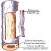
The system of hollow elastic tubes that differ in structure, diameter, mechanical properties are vessels. According to the type of movement, they are divided into arteries (correctly - from the heart to the organs), veins (to the heart from the organs). Capillaries (pictured) are small anatomical blood vessels that permeate all cells and tissues of the body. The hollow veins are distinguished by thin venous walls, a reduced amount of muscle, elastic tissue.

Anatomy and physiology of the heart
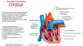
A hollow muscular organ, rhythmically contracting, responsible for the continuity of blood flow through the vessels, is called the heart. The anatomy of the human cardiovascular system calls it the main component. The size of the heart is about the size of a fist, weight is 500 g. The strong organ consists of four chambers, divided by a septum into the right and left halves: the lower ones are the ventricles, the upper chambers are the atria. Each ventricle with the atrium of one side is connected by an atrioventricular opening, an opening, closing valve.
Functions
The main and most important functions of the cardiovascular system are to provide organs with nutrients, biologically active components, oxygen, and energy. Decay products are removed with the blood. The most important function of the heart is the pumping of blood from the veins into the arteries, the transfer of kinetic energy to the blood. It is also called a pump because of physiology. The heart is distinguished by its high productivity, the speed of the processes, the safety margin and stable tissue renewal, forms the nervous regulation of the vascular circles.
Circles of blood circulation
Man and all vertebrates have a closed circulatory system, consisting of vessels of the small, large circle of blood circulation with central nerve impulses. Small or respiratory serves to carry blood from the heart to the lungs, in the opposite direction. It starts from the right ventricle, the pulmonary trunk, ends with the left atrium with the flowing pulmonary arteries, veins. The large one is used to connect the heart with other parts of the body. It begins with the aorta of the left ventricle, forms the veins of the right atrium.
In small, due to venous pressure, the blood is saturated with oxygen, the removal of carbon dioxide by pulmonary capillaries - the smallest vessels. Additionally, the following cardiovascular channels of the circulatory system are distinguished:
- placental - in the fetus in the uterus;
- heart - part of the great circle;
- willis - arteries of the vertebrates, internal carotid arteries at the base of the brain, needed to compensate for the lack of blood supply to the organs.

Cardiovascular diseases
The main organs of the cardiovascular system are prone to various diseases. The most common cardiovascular pathologies are called:
To cure cardiovascular diseases, medications prescribed by a doctor are used, taken in a certain course. They help to normalize the system, eliminate failures. Common drugs and procedures:
- Nitrates - for vasodilation, reduction of ischemia, angina pectoris, disease prevention. Refers to Nitrosprey, Nitromint, Nitroglycerin.
- Antiplatelet agents - for ischemia, defect to reduce platelet aggregation. Low-dose aspirin, Ticlopidine are included.
- Anticoagulants - against excess blood clotting. Direct Heparin, Enoxaparin and indirect Warfarin are used for myocardial infarction, angina pectoris, atrial fibrillation.
- Calcium channel blockers - Verapamil, Nifedipine are used for arrhythmias, tachycardia, pulmonary hypertension.
- Diuretics - Furosemide, Indapamide are used for congestive heart failure, hypertension, remove fluid.
- Lipid-lowering drugs - statins (Atorvastatin) and fibrates (Fenofibrate) lower blood cholesterol levels, prevent atherosclerosis.
- Antiarrhythmic medicines, cardiac glycosides - for circulatory failure. Strengthens the strength and efficiency of heart contractions.
The cardiovascular system - an organ system that ensures blood circulation in humans and animals. Thanks to blood circulation, oxygen and nutrients are delivered to the organs and tissues of the body, while carbon dioxide, other metabolic products and waste products are excreted.
The circulation of blood in the cardiovascular system in vertebrates and humans is supplemented by lymph outflow from the organs and tissues of the body through the system of vessels, nodes and ducts of the lymphatic system, flowing into the venous system at the confluence of the subclavian veins.
The heart is part of the cardiovascular system - an organ that makes blood move, forcing it into blood vessels - hollow tubes of various sizes through which it circulates.
All functions of the circulatory system are strictly coordinated due to neuro-reflex regulation, which allows you to maintain homeostasis in the constantly changing conditions of the external and internal environment.
Blood vessels - these are hollow tubes through which blood flows. The vessels that carry blood from the heart to the organs are called arteries, and from organs to the heart are called veins. There is no gas exchange and diffusion of nutrients in the arteries and veins, it is just a delivery route. As the blood vessels move away from the heart, they become smaller.
Among the vessels of the circulatory system, arteries, arterioles, precapillaries, capillaries, postcapillaries, venules, veins, and arterio-venous anastomoses are distinguished.
The exchange of substances between blood and interstitial fluid occurs through the permeable wall of capillaries - small vessels that connect the arterial and venous systems. In one minute, about 60 liters of liquid seeps through the walls of all human capillaries.
There is a microvasculature between the arteries and veins, which forms the peripheral part of the cardiovascular system. The microcirculatory bed is a system of small vessels, including arterioles, capillaries, venules, and arteriovenular anastomoses. It is here that the processes of exchange between blood and tissues take place.
Although blood with oxygen and nutrients for cells is called arterial, and blood with carbon dioxide and metabolic products of cells is venous, it is not at all necessary that arterial blood flows through the arteries, and venous blood flows through the veins. It depends on the circulation.
The vascular system can be closed - when the blood inside the vessels moves in a circle, and open - when the lumen of the vessels opens freely into the intercellular space and blood flows there, mixing with the extracellular fluid.
A heart(lat. cor, Greek. καρδιά ) is a hollow muscular organ that pumps blood through the vessels with a sequence of contractions and relaxation. Depending on the species, the interior can be divided by partitions into two, three or four chambers. In mammals and birds, the heart is four-chambered. At the same time, they are distinguished (by blood flow): the right atrium, right ventricle, left atrium and left ventricle.
The wall has three layers: the inner one is the endocardium (its outgrowths form the valves), the middle one is the myocardium (cardiac muscle, contraction does not occur arbitrarily, the atria and ventricles do not connect with each other), the outer one is the epicardium (covers the surface of the heart, serves as an inner layer of the pericardial serous membrane - pericardium).
The anatomy of the heart largely determines the degree of basal metabolism, dividing animals into warm-blooded and cold-blooded.
The nerve centers that regulate the activity of the heart are located in the medulla oblongata. These centers receive impulses that signal the needs of certain organs. In turn, the medulla oblongata sends signals to the heart: to strengthen or weaken cardiac activity. The need of organs for blood flow is recorded by two types of receptors: stretch receptors and chemoreceptors.
During the work of the heart, sounds arise - tones:
1. Systolic - low, long-term (oscillation of the valves, two- and tricuspid valves slam, oscillations pull the tendon threads).
2. Diastolic - high, short (the semilunar valves of the aorta and pulmonary trunk are closed).
The heart beats rhythmically at rest with a frequency of 60-70 beats per minute. Frequency below 60 -bradycardia, above 90 - tachycardia.
Contraction of the muscles of the heart is characterized by the contraction time: atria - 0.1 seconds, contraction of the ventricles - 0.3 seconds, pause - 0.4 seconds.
Circles of human circulation
Where the vascular system is closed, it formscircle of blood circulation ... Man and all vertebrates have several circles of blood circulation, exchanging blood with each other only in the heart. The circle of blood circulation consists of two successively connected circles (loops), starting with the ventricles of the heart and flowing into the atria.
The human cardiovascular system forms two circles of blood circulation: large and small.
· A large circle of blood circulation begins in the left ventricle and ends in the right atrium, where they flowhollow veins
· Small circle of blood circulation begins in the right ventricle from whichpulmonary trunk , and ends in the left atrium, into whichpulmonary veins
The systemic circulation provides blood to all organs and tissues.
The small circle of blood circulation is limited by blood circulation inlungs, blood is enriched with oxygen and carbon dioxide is removed.
Depending on the physiological state of the body, as well as practical feasibility, sometimes additional circles of blood circulation are distinguished:
· placental - exists in a fetus inuterus
· cardiac - is part of the systemic circulation
· willis - an arterial ring formed by the arteries of the basin of the vertebral and internal carotid arteries, located at the base of the brain, helps to compensate for insufficient blood supply