Who discovered the existence of cells in 1665. The story of the discovery of the cell. Creation of a cell theory. Cellular theory in its modern form includes three main provisions
- an elementary structural and functional unit of all living organisms It can exist as a separate organism (bacteria, protozoa, algae, fungi), and in the tissues of multicellular animals, plants and fungi.
The history of the study of the cell. Cell theory.
The vital activity of organisms at the cellular level is studied by the science of cytology or cell biology. The emergence of cytology as a science is closely related to the creation of cell theory, the broadest and most fundamental of all biological generalizations.
The history of cell research is inextricably linked with the development of research methods, primarily with the development of microscopic technology. For the first time the microscope was used for research of plant and animal tissues by the English physicist and botanist Robert Hooke (1665). Studying a cut of an elderberry core cork, he discovered separate cavities - cells or cells.
In 1674, the famous Dutch researcher Anthony de Leeuwenhoek improved the microscope (magnified 270 times) and discovered unicellular organisms in a drop of water. He found bacteria in dental plaque, discovered and described erythrocytes, spermatozoa, and described the structure of the heart muscle from animal tissues.
- 1827 - our compatriot K. Baer discovered an egg.
- 1831 - English botanist Robert Brown described the nucleus in plant cells.
- 1838 - German botanist Matthias Schleiden put forward the idea of the identity of plant cells in terms of their development.
- 1839 - German zoologist Theodor Schwann made the final generalization that plant and animal cells have a common structure. In his work "Microscopic studies on the correspondence in the structure and growth of animals and plants," he formulated the cellular theory, according to which cells are the structural and functional basis of living organisms.
- 1858 - German pathologist Rudolf Virchow applied the cellular theory to pathology and supplemented it with important provisions:
1) a new cell can arise only from the previous cell;
2) human diseases are based on a violation of the structure of cells.
Cellular theory in its modern form includes three main provisions:
1) a cell is an elementary structural, functional and genetic unit of all living things - the primary source of life.
2) new cells are formed as a result of division of the previous ones; a cell is an elementary unit of the development of living things.
3) the structural and functional units of multicellular organisms are cells.
Cell theory has had a fruitful influence on all areas of biological research.
People learned about the existence of cells after the invention of the microscope. The very first primitive microscope was invented by the Dutch glass grinder Z. Jansen (1590) by connecting two lenses together.
English physicist and botanist R. Hooke, having examined the cut of cork oak cork, found that it consists of cells, similar to honeycombs, which he called cells (1665). Yes, yes ... this is the same Hooke, after whom the famous physical law is named.
Rice. "A cut of balsa wood from the book by Robert Hooke, 1635-1703"

In 1683, the Dutch researcher A. Van Leeuwenhoek, having improved the microscope, observed living cells and described bacteria for the first time.

Russian scientist Karl Baer discovered a mammalian egg cell in 1827. With this discovery, he confirmed the previously expressed idea of the English physician W. Harvey that all living organisms develop from an egg.
The nucleus was first discovered in plant cells by the English biologist R. Brown (1833).

The works of German scientists, botanist M. Schleiden and zoologist T. Schwann, were of great importance for understanding the role of the cell in living nature. They were the first to formulate cell theory, the main point of which stated that all organisms, including plants and animals, consist of the simplest particles - cells, and each cell is an independent whole. However, in the body, cells work together to form a harmonious unity.
Later in cell theory new discoveries were added. In 1858, the German scientist R. Virchow proved that all cells are formed from other cells by cell division: "every cell is from a cell."
The cell theory served as the basis for the emergence in the 19th century. science of cytology. By the end of the XIX century. due to the complication of microscopic technology, the structural components of cells and the process of their division were discovered and studied. The electron microscope made it possible to study the finest structures of cells. An amazing similarity was found in the fine structure of the cells of representatives of all kingdoms of living nature.
The main provisions of modern cell theory:
- a cell is a structural and functional unit of all living organisms, as well as a unit of development;
- the cells have a membrane structure;
- the nucleus is the main part of the eukaryotic cell;
- cells reproduce only by division;
- the cellular structure of organisms indicates that plants and animals have a single origin.
1. I saw and described plant cells for the first time: R. Virkhov; R. Hooke; K. Baer; A. Levenguk. 2. Improved the microscope and saw unicellular organisms for the first time: M. Schleiden; A. Levenguk; R. Virkhov; R. Hooke.
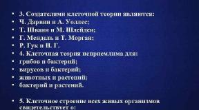
 3. The creators of the cell theory are: C. Darwin and A. Wallace; T. Schwann and M. Schleiden; G. Mendel and T. Morgan; R. Hooke and N. G. 4. The cell theory is unacceptable for: fungi and bacteria; viruses and bacteria; animals and plants; bacteria and plants. 5. The cellular structure of all living organisms testifies to: the unity of the chemical composition; variety of living organisms; the unity of the origin of all living things; the unity of living and inanimate nature


 Prokaryotes are organisms whose cells do not have a nucleus. Prokaryotes (from Latin pro - before, instead of the Greek karion nucleus) - over the kingdom of organisms, which includes the kingdoms of Archaea (Archaebacteria) and Real bacteria (Eubacteria). Bacteria and cyanobacteria themselves are real bacteria (the old name is “blue-green algae”). An analogue of the nucleus is a structure consisting of DNA, proteins and RNA.
 Prokaryotic cells have a surface apparatus and cytoplasm, which contains few organelles and various inclusions. Prokaryotic cells do not have most organelles (mitochondria, plastids, endoplasmic reticulum, Golgi complex, lysosomes, cell center, etc.).
 The sizes of prokaryotes usually range from 0.2 to 30 µm in diameter or length. Sometimes their cells are much larger; thus, some species of the genus Spirochete can reach up to 250 microns in length. The shape of prokaryotic cells is diverse: spherical, rod-shaped, in the form of a comma or spirally twisted filament, etc.
 The structure of the surface apparatus of prokaryotic cells includes a plasma membrane, a cell wall, and sometimes a mucous capsule. In most bacteria, the cell wall consists of a high molecular weight organic compound, murein. This connection forms a mesh structure that stiffens the cell wall.
 In cyanobacteria, the outer layer of the cell wall contains the polysaccharide pectin and special contractile proteins. They provide forms of movement such as sliding or rolling.
 The cell wall often includes a thin layer - the so-called outer membrane, which, like the plasma membrane, contains proteins, phospholipids and other substances. It provides an increased degree of protection for the contents of the cell. The bacterial cell wall has antigenic properties.
 The mucous capsule consists of mucopolysaccharides, proteins or polysaccharides with protein inclusions. It is not very tightly bound to the cell and is easily destroyed by the action of certain compounds. The cell surface of some bacteria is covered with numerous thin filamentous outgrowths. With their help, bacterial cells exchange hereditary information, adhere to each other or attach to the substrate.
 Ribosomes of prokaryotes are smaller than the ribosomes of eukaryotic cells. The plasma membrane can form smooth or folded invaginations into the cytoplasm. The folded membrane invaginations contain respiratory enzymes and ribosomes, and the smooth ones contain photosynthetic pigments.
 In the cells of some bacteria (for example, purple ones), photosynthetic pigments are found in closed sac-like structures formed by invaginations of the plasma membrane. Such bags can be placed singly or collected in piles. Such formations of cyanobacteria are called thylakoids; they contain chlorophyll and are located singly in the surface layer of the cytoplasm.
 Some bacteria and cyanobacteria living in reservoirs or water-filled soil capillaries have special gas vacuoles filled with a gas mixture. By changing their volume, bacteria can move in the water column with minimal energy consumption.
 Many real bacteria have one, several, or many flagella. Flagella can be several times longer than the cell itself, and their diameter is insignificant (10 -25 nm). The flagella of prokaryotes only outwardly resemble the flagella of eukaryotic cells and consist of a single tube formed by a special protein. Cyanobacterial cells lack flagella.
 Features of the vital processes of prokaryotes § Prokaryotic cells can absorb substances with only a small molecular weight. Their entry into the cell is provided by the mechanisms of diffusion and active transport. § Prokaryotic cells reproduce exclusively asexually: by dividing in two, occasionally by budding. Before division, the hereditary material of the cell (DNA molecule) is doubled.
 Transfer of unfavorable conditions by prokaryotes When unfavorable conditions occur, sporulation occurs in some prokaryotes. Some prokaryotes are capable of encysting (from Latin in - in, inside and Greek cyst - bubble). In this case, the entire cell is covered with a dense membrane. Prokaryotic cysts are resistant to radiation, drying, but, unlike spores, they are unable to withstand exposure to high temperatures. In addition to experiencing adverse conditions, spores and cysts ensure the spread of prokaryotes using water, wind, or other organisms.
 Let's draw conclusions § Prokaryotic cells do not have a nucleus and many organelles (mitochondria, plastids, endoplasmic reticulum, Golgi complex, lysosomes, cell center, etc.). Prokaryotes are unicellular or colonial organisms. § The surface apparatus of prokaryotic cells includes a plasma membrane, a cell wall, and sometimes a mucous capsule located above it. The cell wall of most bacteria contains a high molecular weight organic compound, murein, which makes it hard. § The cytoplasm of prokaryotes contains small ribosomes and various inclusions. The plasma membrane can form smooth or folded invaginations into the cytoplasm. Respiratory enzymes and ribosomes are located on folded membrane invaginations, on
 Let's draw conclusions § In prokaryotic cells there are one or two nuclear zones of the nucleoid, where the hereditary material is located - a circular DNA molecule. § The cells of some bacteria have organelles that move one, several or many flagella. § Prokaryotic cells multiply by dividing in two, sometimes by budding. For some species, the known process of conjugation, during which cells exchange DNA molecules. Spores and cysts provide prokaryotes with the experience of unfavorable conditions and spread in the biosphere.
Cytology ("cytos" - cell, cell) is the science of a cell. Modern cytology studies: the structure of cells, their formation as elementary living systems, studies the formation of individual cellular components, the processes of cell reproduction, repair, adaptation to environmental conditions and other processes. In other words, modern cytology is the physiology of the cell.
The development of the theory of the cell is closely connected with the invention of the microscope (from the Greek "micro" - small, "scopo" - I am considering). This is due to the fact that the human eye is unable to distinguish objects with dimensions less than 0.1 mm, which is 100 micrometers (abbreviated microns or microns). The sizes of cells (and even more so, of intracellular structures) are significantly smaller.
For example, the diameter of an animal cell usually does not exceed 20 µm, a plant cell - 50 µm, and the chloroplast length of a flowering plant does not exceed 10 µm. With the help of a light microscope, objects with a diameter of tenths of a micron can be distinguished.
The first microscope was designed in 1610 by Galileo and consisted of a combination of lenses in a lead tube (fig. 1.1). And before this discovery in 1590, the Dutch master Jansens were engaged in glass making.
Rice. 1.1. Galileo Galilei (1564-1642)
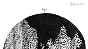
For the first time the microscope was used for research by the English physicist and natural scientist R. Hooke (fig. 1.2, 1.4). In 1665, he first described the cellular structure of the cork and introduced the term "cell" (fig. 1.3). R. Hooke made the first attempt to count the number of cells in a certain volume of the cork.
He formulated the concept of a cell as a cell completely closed from all sides and established the fact of the cellular structure of plant tissues. These two main conclusions determined the direction of further research in this area.

Rice. 1.2. Robert Hooke (1635-1703)

Rice. 1.3. Cork cells studied by Robert Hooke

Rice. 1.4. Microscope by Robert Hooke
In 1674, the Dutch merchant Antonio van Leeuwenhoek, using a microscope, first saw in a drop of water "animals" - moving living organisms (unicellular organisms, blood cells, sperm) and reported this to the scientific community (fig. 1.5, 1.6)... The descriptions of these "animalalcus" made the Dutchman world famous, aroused interest in the study of the living microworld.

Rice. 1.5. Antonio van Leeuwenhoek (1632-1723)

Rice. 1.6. Microscope by Antonio van Leeuwenhoek
In 1693, during the stay of Peter I in Delphi, A. Levenguk demonstrated to him how the blood moves in the fin of a fish. These demonstrations made such a great impression on Peter I that, after returning to Russia, he created a workshop for optical instruments. In 1725, the St. Petersburg Academy of Sciences was organized.
The talented masters I.E. Belyaev, I.P. Kulibin made microscopes (fig. 1.7, 1.8, 1.9), in the design of which academicians L. Euler, F. Epinus took part.

Rice. 1.7. I.P. Kulibin (1735-1818)

Rice. 1.8. I.E. Belyaev

Rice. 1.9. Microscopes made by Russian craftsmen
In the years 1671-1679. Italian biologist and physician Marcello Malpighi gave the first systematic description of the microstructure of plant organs, which laid the foundation for plant anatomy (fig. 1.10).

Rice. 1.10. Marcello Malpighi (1628-1694)
In the years 1671-1682. the Englishman Nehemiah Grew described in detail the microstructure of plants; introduced the term "tissue" to denote the concept of a collection of "bubbles", or "sacs" (fig. 1.11)... Both of these researchers (they worked independently of each other) gave amazingly accurate descriptions and drawings. They came to the same conclusion regarding the universality of the construction of plant tissue from bubbles.

Rice. 1.11. Nehemiah Gru (1641-1712)
In the 20s of the XIX century. the most significant works in the field of the study of plant and animal tissues belonged to the French scientists Henri Dutrochet (1824), Francois Raspail (1827), Pierre Turpin (1829). They proved that cells (sacs, vesicles) are the elementary structures of all plant and animal tissues. These studies paved the way for the discovery of cell theory.
One of the founders of embryology and comparative anatomy, academician of the St. Petersburg Academy of Sciences Karl Maksimovich Baer showed that a cell is a unit not only of structure, but also of the development of organisms (fig. 1.12).

Rice. 1.12. K.M. Baer (1792-1876)
In 1759, the German anatomist and physiologist Caspar Friedrich Wolf proved that a cell is a unit of growth. (fig. 1.13).

Rice. 1.13. K.F. Wolf (1733-1794)
1830s Czech physiologist and anatomist J.E. Purkine (fig. 1.14), German biologist I.P. Müller proved that cellular organization is universal for all types of tissues.

Rice. 1.14. Ya.E. Purkine (1787-1869)
In 1833 the British botanist R. Brown (fig. 1.15) described the nucleus of a plant cell.

Rice. 1.15. Robert Brown (1773-1858)
In 1837 Matthias Jakob Schleiden (fig. 1.16) proposed a new theory of the formation of plant cells, recognizing the decisive role in this process of the cell nucleus. In 1842, he first discovered the nucleoli in the nucleus.
According to modern concepts, Schleiden's specific studies contained a number of errors: in particular, Schleiden believed that cells can arise from a structureless substance, and a plant embryo can develop from a pollen tube (the hypothesis of spontaneous generation of life).

Rice. 1.16. Matthias Jakob Schleiden (1804-1881)
German cytologist, histologist and physiologist Theodor Schwann (fig. 1.17) got acquainted with the works of the German botanist M. Schleiden, which described the role of the nucleus in the plant cell. Comparing these works with his own observations, Schwann developed his own principles of cellular structure and development of living organisms.
In 1838, Schwann published three preliminary reports of the cell theory, and in 1839 - the work "Microscopic studies on the correspondence in the structure and growth of animals and plants", where he published the basic principles of the theory of the cellular structure of living organisms.
F. Engels argued that the creation of the cellular theory was one of the three greatest discoveries in natural science in the 19th century, along with the law of energy conversion and evolutionary theory.

Rice. 1.17. Theodor Schwann (1810-1882)
In 1834-1847. Professor of the Medical-Surgical Academy in St. Petersburg P.F. Goryaninov (fig. 1.18) formulated the principle according to which the cell is a universal model of the organization of living beings.
Goryaninov divided the world of living beings into two kingdoms: the formless kingdom, or molecular, and organic, or cellular. He wrote that "... the organic world is, first of all, the cellular kingdom ...". He noted in his studies that all animals and plants consist of interconnected cells, which he called bubbles, that is, he expressed an opinion about the general plan of the structure of plants and animals.

Rice. 1.18. P.F. Goryaninov (1796-1865)
In the history of the development of cell theory, two stages can be distinguished:
1) the period of accumulation of observations on the structure of various unicellular and multicellular organisms of plants and animals (about 300 years);
2) the period of generalization of the available data in 1838 and the formulation of the postulates of the cell theory;
The first person to see the cells was an English scientist Robert Hooke(known to us thanks to Hooke's law). V 1665 year trying to understand why corkwood swims so well, Hooke began to examine the thin sections of the cork with the help of his improved microscope... He found that the cork was divided into many tiny cells that reminded him of monastery cells, and he called these cells cells (in English, cell means “cell, cell, cage”). V 1675 year italian doctor M. Malpighi and in 1682 year- English botanist N. Grew confirmed the cellular structure of plants. The cell began to be spoken of as a "bubble filled with nutritious juice." V 1674 year Dutch master Antony van Leeuwenhoek(Anton van Leeuwenhoek, 1632
-1723
) with the help of a microscope for the first time I saw in a drop of water "animals" - moving living organisms ( ciliates, amoeba, bacteria). Also, Leeuwenhoek was the first to observe animal cells - erythrocytes and sperm... Thus, by the beginning of the 18th century, scientists knew that under high magnification, plants have a cellular structure, and they saw some organisms, which were later called unicellular. V 1802
-1808 years French explorer Charles-Francois Mirbel found that all plants are composed of tissues formed by cells. J. B. Lamarck v 1809 year extended Mirbel's idea of the cellular structure to animal organisms. In 1825 a Czech scientist J. Purkine discovered the nucleus of the egg cell of birds, and in 1839
introduced the term “ protoplasm". In 1831, an English botanist R. Brown first described the nucleus of a plant cell, and in 1833 year found that the nucleus is an indispensable organoid of the plant cell. Since then, the main thing in the organization of cells is not the membrane, but the content.
Cell theory the structure of organisms was formed in 1839 year German zoologist T. Schwann and M. Schleiden and included three provisions. In 1858 Rudolf Virchow supplemented it with one more provision, but his ideas contained a number of mistakes: for example, he assumed that the cells are weakly connected to each other and each exists "by itself." Only later was it possible to prove the integrity of the cellular system.
V 1878 year Russian scientists I. D. Chistyakov open mitosis in plant cells; v 1878 year V. Flemming and P. I. Peremezhko reveal mitosis in animals. V 1882 year V. Flemming observes meiosis in animal cells, and in 1888 year E Strasburger - in vegetable.
18. Cell theory- one of the generally recognized biological generalizations that affirm the unity of the principle of the structure and development of the world plants, animals and other living organisms with cellular structure, in which the cell is considered as a common structural element of living organisms.