Becker's progressive muscular dystrophy. Becker's myopathy. Don't waste time
Duchenne and Becker myodystrophy is an X-linked recessive disorder characterized by progressive proximal muscle weakness due to degeneration of muscle fibers. Becker's myodystrophy is characterized by a later onset and less severe manifestations.
The diagnosis is suggested clinically and confirmed by determining the protein (dystrophin), which is the product of the mutated gene. Treatment focuses on maintaining function with physical therapy and the use of braces and orthopedic devices; some patients with a marked decrease in function are prescribed prednisone.
What causes Duchenne and Becker muscular dystrophy?
Duchenne and Becker dystrophies are caused by mutations at the Xp21 locus. In Duchenne dystrophy, the mutation results in the absence of dystrophin, a protein in the cell wall membrane. With Becker's dystrophy, as a result of mutation, an abnormal dystrophy or an insufficient amount is synthesized. Duchenne myodystrophy occurs in 1/3000 live-born boys; Becker's dystrophy - in 1/30 000 live-born boys.
Duchenne and Becker muscular dystrophy symptoms
Duchenne myodystrophy usually appears between two and three years of age. Weakness of the proximal muscles develops, usually beginning in the lower extremities. Children develop a waddling gait, a sock-supported gait, and lordosis. Such children often fall, it is difficult for them to run, jump, climb stairs and off the floor. The disease Duchenne myodystrophy is steadily progressing, flexion contractures of the joints and scoliosis occur. Dense pseudohypertrophy develops (fatty and fibrous replacement of certain enlarged muscle groups, especially the calf muscles). Most patients are wheelchair-bound by age 12 and die of respiratory complications by age 20. Heart damage is usually asymptomatic, despite the fact that 90% of patients have ECG changes. A third of patients have a mild non-progressive decrease in intelligence, to a greater extent with a violation of verbal rather than non-verbal tests.
Becker's myodystrophy manifests itself clinically much later, and its symptoms are less pronounced. Patients are usually able to walk until at least 15 years of age, and many remain walking into adulthood. Most affected patients live for more than 30-40 years.
Diagnostics of the Duchenne and Becker muscular dystrophy
The diagnosis is suspected on the basis of characteristic clinical manifestations, age of onset, family history indicating an X-linked type of inheritance. Signs of myopathy are found on electromyography (rapidly evoked, short, low-amplitude motor potentials) and muscle biopsy (necrosis and marked differences in muscle fiber size). Creatine kinase levels can increase up to 100 times the normal level.
The diagnosis is confirmed by dystrophin immunostaining. Dystrophy is not defined in patients with Duchenne dystrophy; in patients with Becker's dystrophy, the dystrophy is usually abnormal (with a lower molecular weight) or detected at lower concentrations. DNA analysis of peripheral blood leukocytes to detect mutations can also confirm the diagnosis when abnormalities in the dystrophin gene are detected (deletions and duplications in about 65% and point mutations in about 25% of patients).
Carrier detection and prenatal diagnosis are possible using conventional methods (pedigree analysis, creatine kinase determination, fetal sex determination) in combination with DNA analysis and muscle tissue immunostaining with antibodies to dystrophin.
, , , , , , , , [
A variant of hereditary X-linked myodystrophy, characterized by a slower and more benign course. The disease is characterized by progressively worsening and spreading muscle weakness, hypotension, and atrophy, initially occurring in the muscles of the thighs and pelvic girdle. Diagnostic search includes neurological examination, consultation of a geneticist and cardiologist, neurophysiological testing of the neuromuscular apparatus, DNA diagnostics, muscle biopsy with morphological, immunological and histochemical study of the obtained samples. Treatment is symptomatic and, unfortunately, ineffective. The progression of the disease leads to the loss of patients' ability to move independently by the age of 40.

General information
Becker's progressive muscular dystrophy was first described in 1955 as a benign variant of the course of Duchenne muscular dystrophy. Subsequently, numerous studies in the field of clinical neurology, genetics and biochemistry revealed significant differences in the nature of the course, biochemical and morphological basis of these diseases. As a result, the clinical form of Becker was identified as an independent nosology.
Becker muscular dystrophy belongs to the group of myopathies (myodystrophies) - diseases arising from disorders of the structure and metabolism of muscle tissue and manifested by muscle weakness. The pathology is inherited recessively linked to the X chromosome, therefore only males are ill. The frequency of occurrence is 1 newborn per 20 thousand children.
Causes of occurrence
At the heart of the disease is a mutation in the gene responsible for encoding the dystrophin protein. Approximately 30% of the total number of cases of Becker muscular dystrophy occurs in the so-called. "Fresh" mutations. The gene is located at 21 loci (in the region Xp21.2 – p21.1) of the short arm of the X chromosome. Approximately 65-70% of patients have large deletions of this region, 5% have duplications, and the rest have point mutations. These structural rearrangements of the gene do not entail a complete cessation of dystrophin synthesis, as in Duchenne dystrophy, but potentiate the synthesis of an abnormal truncated protein, to some extent capable of performing its functions. This explains the more benign nature of Becker's dystrophy in comparison with the Duchenne variant.
Normally, the protein dystrophin maintains the integrity of the sarcolemma - the membrane of myocytes (muscle fibers), provides the elasticity and stability of myofibrils when muscle contraction... The inability of abnormal dystrophin to adequately perform these functions leads to disruption of the integrity of the membranes of muscle fibers. As a consequence of this, degenerative changes in the cytoplasmic components of the latter and increased transport of potassium ions into the myocytes occur. The result of such biochemical and morphological changes is the death of myofibrils and the destruction of muscle fibers. In place of the dead myocytes, the formation of connective tissue, which causes the phenomenon of pseudohypertrophy - an increase in muscle volume and density with a sharp decrease in its contractility.
Symptoms
Becker's progressive muscular dystrophy usually manifests itself between 10 and 15 years of age, in some cases earlier. The initial signs of the disease are excessive fatigue and muscle weakness in the pelvic girdle and lower limbs... In a number of patients, the first manifestations are periodic painful muscle cramps (cramps) localized in the legs. Muscle weakness makes it difficult to climb stairs, if necessary, get up from a sitting position. Over time, a waddling "duck" gait is formed. In order to get up, the patient is forced to use auxiliary myopathic techniques - to lean with his hands on nearby pieces of furniture or, in the absence of such, use his own body as a support (Govers symptom).
Like other hereditary myopathies, Becker's disease is characterized by symmetrically developing muscle atrophy. First of all, the muscles of the thigh and pelvic girdle are affected, then the process extends to the muscles of the shoulder girdle and proximal muscles of the arms. At the onset of the disease, pseudohypertrophies are formed, most pronounced in the gastrocnemius, deltoid, triceps and quadriceps muscles. As myodystrophy progresses, they transform into muscle wasting.
The clinical picture of Becker muscular dystrophy is in many respects similar to Duchenne muscular dystrophy. Aggravation of muscle weakness over time leads to immobility of the patient and the formation of joint contractures. However, the development of a dystrophic process in muscle tissue with Becker's dystrophy is much slower, which causes prolonged motor activity of patients. On average, patients retain the ability to move independently until 35-40 years of age. In addition, Becker's dystrophy is not accompanied by oligophrenia, severe curvature of the spine and other skeletal deformities. Possible cardiomyopathy of dilated or hypertrophic type, blockade of the bundle of His, but cardiovascular disorders are moderate. There may be a decrease in libido, gynecomastia, testicular atrophy, impotence.
Diagnostics
Becker's progressive muscular dystrophy is diagnosed by a neurologist based on history, clinical data, additional examinations, and genetic testing. In neurological status, there is a decrease in muscle strength and a moderate decrease muscle tone in the proximal extremities, loss of knee reflexes with a symmetrical decrease in tendon reflexes of the distal legs and upper extremities, complete preservation of sensitivity.
Among clinical analyzes highest value has a biochemical blood test, which reveals a multiple increase in the level of CPK. Electroneurography data allow to exclude damage to nerve fibers, electromyography indicates the primary muscular type of damage. A muscle biopsy is done only after a negative genetic test. Morphological study of the obtained material determines diffuse variability, dystrophic and necrotic changes in muscle fibers, proliferation of connective tissue. Special immunostaining of samples is carried out, followed by determination of the presence of dystrophin in them.
To confirm the diagnosis of Becker muscular dystrophy, a geneticist's consultation with DNA analysis allows. Identification of duplications or deletions in the Xp21 gene makes it possible to establish an accurate diagnosis. A negative DNA test result does not indicate the absence of pathology, since point mutations may occur, the search for which is a complex and more expensive procedure.
In order to identify cardiac pathology, electrocardiography, Echo-KG, consultation with a cardiologist are prescribed. Cardiological examination can detect a violation of intraventricular conduction, AV block, ventricular dilatation, hypertrophic changes in the myocardium, cardiomyopathy, heart failure.
Differential diagnosis is carried out with progressive Dreyfus muscular dystrophy, Duchenne muscular dystrophy, Erb-Roth muscular dystrophy, metabolic myopathy, polymyositis and dermatomyositis, inflammatory myopathy, spinal amyotrophy, hereditary polyneuropathy.
Prenatal diagnosis is recommended when the mother is a carrier of the pathogenic gene. If the child is male, then the probability of developing the disease is 50%. Chorionic biopsy can be performed at 11-14 weeks. pregnancy, amniocentesis - after the 15th week, cord blood sampling (cordocentesis) - for more than 18 weeks.
Treatment
On the the present stage several groups of scientists are conducting persistent research in the field of search effective methods treatment of progressive muscular dystrophies. Currently, patients receive mainly metabolic and symptomatic therapy. Various treatment regimens have been developed to improve the patient's motor abilities and somewhat slow down the progression of the disease. Patients are prescribed actoprotectors (ethylthiobenzimidazole), neostigmine, ATP, anabolic steroid(methyandrostenediol), cardiac drugs. On the issue of long-term therapy with glucocorticoids (prednisolone), clinicians have different opinions. Some believe that such treatment inhibits the progression of myodystrophy, others reject this assumption.
Observations have shown that bed rest aggravates muscle weakness. Therefore, patients are advised to moderate physical activity swimming. The maintenance of muscle elasticity and strength, as well as the prevention of contractures, is carried out by means of massage, physiotherapy and remedial gymnastics. Surgical treatment of contractures is performed according to indications. The use of various orthopedic aids (walkers, wheelchairs, leg braces, exoskeletons) allows you to expand the movement capabilities of patients and their ability to self-care. Surgical treatment of contractures is performed according to indications.
Forecast and prevention
Becker's progressive muscular dystrophy has a poor prognosis. Although immobility in patients occurs much later than in Duchenne dystrophy, ultimately damage to the heart muscle and respiratory muscles leads to the death of patients from heart or respiratory failure. Thoughtful care, adequate therapy, ventilation support for breathing, the use of orthopedic aids can only increase the duration and improve the patient's quality of life. Prevention consists in preventing the birth of a child with pathology through genetic counseling of future parents and prenatal diagnosis.
Etiology and incidence of Duchenne muscular dystrophy... Duchenne muscular dystrophy (MIM # 310200) is a pan-ethnic X-linked progressive myopathy caused by mutations in the DMD gene. The incidence is approximately 1 in 3500 male newborns.
The pathogenesis of Duchenne muscular dystrophy... The DMD gene encodes dystrophies, an intracellular protein expressed predominantly in smooth, skeletal and cardiac muscle, as well as in some neurons in the brain. In skeletal muscles, dystrophy is part of a large complex of sarcolemma-associated proteins that provide sarcolemma resistance.
Mutations in gene DMD that cause Duchenne muscular dystrophy include large deletions (60-65%), large duplications (5-10%), and small deletions, insertions or substitutions of nucleotides (25-30%). The largest deletions occur in one of two hotspots. Nucleotide substitutions occur throughout the gene, predominantly in CpG dinucleotides.
De novo mutations occur with a comparable frequency during ovogenesis and spermatogenesis; the largest de novo deletions occur during ovogenesis, while most de novo nucleotide substitutions occur during spermatogenesis.
Mutations causing the phenotypic absence of dystrophin lead to more severe muscle damage than mutant DMD alleles expressing partially functional dystrophies. No correlation was found between genotype and phenotype for intellectual decline.
Phenotype and development of Duchenne muscular dystrophy
Men with Duchenne muscular dystrophy... Myodystrophy is a progressive myopathy that leads to muscle degeneration and weakness. Starting with the muscles of the hip girdle and the flexors of the neck, muscle weakness progressively affects the shoulder girdle and distal muscles of the limbs and trunk. Although occasionally, patients are accidentally diagnosed during the neonatal period due to hypotension or developmental delay, usually sick boys are diagnosed at the age of 3 to 5 years with the appearance of gait anomalies.
By the age of 5, most affected children use Govers' techniques and have pseudohypertrophy of the lower leg muscles, i.e. an increase in the legs due to the replacement of muscles with adipose and connective tissue. By the age of 12, the majority of patients are immobilized in wheelchair and have contractures and scoliosis. Most patients die from impaired pulmonary function and pneumonia; the average age of death is 18 years.
Almost 95% of patients Duchenne muscular dystrophy have certain cardiac abnormalities (dilated cardiomyopathy or electrocardiographic abnormalities), and 84% have visible lesions of the heart muscle at autopsy. Chronic heart disease occurs in almost 50% of patients, occasionally heart failure causes them complaints. Although dystrophy is also present in smooth muscle, smooth muscle complications are rare and include gastric dilatation, volvulus, and bladder hypotension.
Sick Duchenne muscular dystrophy have IQs about 1 standard deviation lower than normal, and almost a third have some degree mental retardation... The reasons for this have not been established.
Women with Duchenne muscular dystrophy
Age of onset and severity Duchenne muscular dystrophy in women, depend on the degree of displacement of the X-chromosome inactivation. If the X chromosome carrying the mutant DMD allele is active in most cells, the woman will develop signs of Duchenne muscular dystrophy; if the X chromosome carrying the normal DMD allele is predominantly active, women have only a few or no symptoms of the disease.
Whether they have clinical symptoms skeletal muscle weakness, female carriers have abnormalities in cardiac muscle function, such as dilated cardiomyopathy, left ventricular dilatation, and electrocardiographic changes.
Features of phenotypic manifestations of Duchenne dystrophy:
Age of onset: childhood
Muscle weakness
Leg hypertrophy
Minor intellectual disability
High serum creatine kinase
Duchenne muscular dystrophy treatment
Duchenne muscular dystrophy diagnosis based on family history and DNA analysis or muscle biopsy with immunohistochemistry for dystrophin.
Currently cure for Duchenne muscular dystrophy not possible, although improved symptomatic treatment increased the average life span from late childhood to early adulthood. The goals of therapy are to slow the progression of disease, provide mobility, prevent or correct contractures and scoliosis, control body weight, and improve lung and heart function.
Glucocorticoid therapy can slow the progression of the disease for several years. Several experimental treatments are being investigated, including gene transfer. Most patients also need extensive counseling as they deal with the psychological effects of chronic fatal illness.
Risk of Inheriting Duchenne Muscular Dystrophy
The third part of mothers who gave birth to a single patient son are themselves carriers of mutations in the DMD gene. Nevertheless, the determination of the carriage remains a difficult task, since currently available molecular methods do not detect small mutations such as single nucleotide substitutions. Determination of the risk of carriage in families without a found deletion or duplication is based on linkage assays, a series of serum creatine casase assays, and mosaic dystrophin expression in muscle biopsy samples (due to accidental X-chromosome inactivation). When counseling for a recurrence risk assessment, the high incidence of germ cell mosaicism (approximately 14%) should be taken into account.
If the mother is a carrier, everyone son has a 50% risk of developing Duchenne muscular dystrophy, and each daughter has a 50% risk of inheriting the DMD mutation. Reflecting the random nature of the X chromosome inactivation, daughters who inherit a mutation in the DMD gene have a low risk of Duchenne muscular dystrophy; however, for reasons completely incomprehensible, the risk of heart abnormalities can be as high as 50-60%. If the mother is not DNA-tested, she has an approximately 7% risk of having a boy with Duchenne muscular dystrophy due to sexual mosaicism. Genetic counseling and possibly prenatal diagnosis are indicated for these mothers.
An example of Duchenne muscular dystrophy... AI, a 7-year-old boy, is being examined for mild developmental delay. He has difficulty climbing stairs, running, reduced strength and endurance with intense physical activity... His parents, two brothers and a sister are completely healthy; other family members have no similar complaints. Examination revealed difficulty in jumping, Govers' techniques (a sequence of movements that facilitate getting up from the floor), weakness of the proximal muscles, waddling ("duck") gait, induration of the Achilles tendons and markedly hypertrophied lower leg muscles. The serum creatine kinase level was 50 times higher than normal.
Insofar as anamnesis and medical examination data, including elevated level creatine kinase, suggested myopathy, the child was sent to the neurogenetics clinic for further examination. Muscle biopsy results showed a pronounced change in the size of muscle fibers, fiber necrosis, proliferation of adipose and connective tissue, and no staining for dystrophy. Based on these results, the child was provisionally diagnosed with Duchenne muscular dystrophy and tested for a deletion in the dystrophin gene; it turned out to have a deletion from exons 45 to 48.
Sergei is 19 years old. Diagnosis: Becker muscular dystrophy.
Sergei's muscular dystrophy began to manifest itself at the age of 10. The disease began with general physical weakness. Especially weakness manifested itself in physical education lessons. Then Sergei began to notice that he squatted differently from his classmates. Classmates sat down gently, and Sergei fell on his haunches and held on to his hand so as not to fall. The boy could not get up without the help of his hands.
The young man began to notice that the weakness was symmetrical in both legs. Sergei's parents, when they went to the doctors about their son's weakness, noted that the calf muscles are very dense and voluminous, very similar to bottles. This condition of the calf muscles is called pseudohypertrophy.
Sergei began to show progressive lumbar lordosis, which progressed more and more over the years, a “duck” gait appeared.
When he got up from the floor, Sergei got up, pulling himself up on his arms. He stopped running, as he stumbled all the time, and the muscle weakness increased so much that he had to stop running after a few steps.
It was with such problems that Sergey turned to me with a request to teach his parents to influence the muscles so that they began to work better.
Dr. Nikonov
Sergei and his parents knew that complete immobilization could occur at the age of 25.
The average life expectancy in patients with Becker muscular dystrophy is 40 years. But if the clinical manifestations of myodystrophy begin to appear at the age of 30, then the mobility of patients remains even up to 60 years. This was the case with one of my patients, who came to me at the age of 60 in a semi-mobile state. After carrying out the procedures according to my method, Emine, that is the name of the patient, began to freely rise from the floor without using her hands, and climb quite quickly up the stairs to the 17th floor without using her hands and without a break for rest. For more information on restoring Emine, see the video:
Sergei had an increased serum creatine phosphokinase concentration and myoglobinuria. Since it is difficult to distinguish Becker's muscular dystrophy from Duchenne muscular dystrophy, maintaining the strength of the flexor muscles of the neck makes it possible to differentiate diagnoses.
Sergei retained the strength of the flexor muscles of the neck - he had Becker's muscular dystrophy, while Emine, Nastya and Yakob, my patients, had the muscles of the neck powerless.
Becker muscular dystrophy occurs when the gene that makes the protein dystrophin is damaged. Dystrophin is produced, but in different configurations. The protein dystrophin is the largest protein in the muscle cell. He makes a frame for the cage so that it is hollow inside. The skeleton of the muscle cell consists of the protein dystrophin and resembles a metal mesh with holes. Through these holes, nutrients enter the cell and waste toxins exit. Since the dystrophin mesh is not complete like a brick wall, this mesh can expand and contract. Two motor proteins inside this scaffold - actin and myosin - expand and narrow the mesh. Actin and myosin can move when given energy. The energy for motor proteins is provided by the mitochondria.
Dr. Nikonov
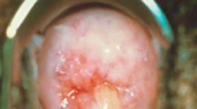
When I looked at the structure of the muscle cell in Becker's muscular dystrophy through an electron microscope, I saw that inside the muscle cell, mitochondria are located not at the motor proteins, but at the periphery of the cell. I also saw that there are a lot of fatty inclusions inside the muscle cell. This means that these fatty inclusions pushed mitochondria away from their jobs and motor proteins cannot work.

This is how muscles look in Becker muscular dystrophy under an electron microscope
A: Normal muscle of a healthy person.
B: Hypertrophied muscle in Becker muscular dystrophy.
Dr. Nikonov
If I act on the muscles with my method, I thought, would the amount of body fat decrease? If the amount of body fat decreases, then is it possible to return the mitochondria back to motor proteins.
As a result of the impact on the muscles with my method, Sergei began to develop Strength in the muscles. He began to walk confidently with his hands down, began to walk at full feet, began to move in space like an ordinary person.

Beginning of procedures to restore the movement of Sergei's muscles
The hardest part is the beginning of the procedures. The first lessons with Sergey were attended by: me, Sergey's parents, my wife Nikonova Lilia Aleksandrovna, our son Stepan (10 years old) and Sergey's sister (10 years old). Our younger assistants were responsible for fixing Sergei's legs. The task for both of them was doable.

Duchenne myodystrophy (DMD)- hereditary disease that begins at the age of 2-5 years and is characterized by progressive muscular weakness, atrophy and pseudohypertrophy proximal muscle, often accompanied by cardiomyopathies and intellectual impairment. In the early stages of the disease, there is increased fatigue when walking, a change in gait ("duck gait"). In this case, there is a gradual degradation of muscle tissue. 95% of patients stop walking at the age of 8-12 years. At the age of 18-20, patients usually die, often from respiratory failure. An allelic form of DMD is distinguished - Becker's muscular dystrophy (BMD, OMIM), which is characterized by similar clinical manifestations, a later onset (at about 10-16 years of age) and a milder course. Such patients often retain the ability to walk for up to 20 years, and some up to 50-60 years, although the same muscles are involved in the pathological process as in DMD. The life expectancy of such patients is reduced slightly.
A biochemical marker of the disease is an increased (100-200) times the level of creatine phosphokinase (KFK) in blood. In carriers of the damaged gene, the CPK level is also slightly increased on average.
The type of inheritance of Duchenne muscular dystrophy is X-linked recessive, i.e. it affects almost exclusively boys, while women with a damaged gene on one of the X chromosomes are carriers of DMD. But in rare cases, girls can also be ill with Duchenne muscular dystrophy. The reasons for this may be the predominant inactivation of the X chromosome with a normal allele in heterozygous carriers of the mutant DMD gene, X autosomal translocation affecting this gene, hemizygosity for the mutant allele, and the presence of phenocopies (diseases associated with disruption of other proteins included in the dystrophin glycoprotein complex ). In approximately 2/3 of cases, the son receives a chromosome with damage from the mother-carrier, in other cases the disease occurs as a result of a de novo mutation in the germ cells of the mother or father, or in the precursors of these cells. Duchenne muscular dystrophy (DMD) occurs in approximately one in 2,500 to 4,000 newborn boys.
The DMD gene, which is responsible for progressive Duchenne / Becker muscular dystrophy (DMD / BMD), is located at the Xp21.2 locus and has a size of 2.6 million bp. and consists of 79 exons. In 60% of cases, mutations leading to DMD / BMD are extended deletions (from one to several tens of exons), in 30% of cases - point mutations, and in 10% of cases - duplications. Due to the presence of so-called "hot spots" of deletions, the amplification of 27 exons and the promoter region of the DMD gene makes it possible to detect approximately 98% of all large deletions. Finding point mutations is difficult due to big size gene and the absence of major mutations.
At the Center for Molecular Genetics, the level of CPK in the blood is measured, as well as direct diagnosis of DMD / BMD, which is a search for large deletions / l duplications in all exons of the DMD gene and the search for "point" mutations of the DMD gene using the NGS (next generation sequensing) method. Research by the NGS method also allows detecting deletions of all exons of the DMD gene in sick boys. Analysis of all exons of a gene allows one to determine the exact exon boundaries of the deletion in the event of its detection, and thus, to establish whether this deletion leads to a shift in the protein reading frame, which in turn is important for predicting the form of the disease - Duchenne or Becker muscular dystrophy. Thus, the combination of various research methods allows detecting almost all mutations of the DMD gene.
The presence of any type of mutation (deletion / duplication in one or several exons, "point" mutations) is a molecular genetic confirmation of the clinical diagnosis of Duchenne / Becker muscular dystrophy and allows prenatal diagnosis in this family.
Attention! To measure the CPK level, blood must be fresh (not frozen)!
In the case of prenatal diagnosis, fetal biomaterial is required, which can be used as chorionic villi (from the 8th to the 12th week of pregnancy), amniotic fluid (from the 16th to 24th week of pregnancy) or umbilical cord blood (from the 22nd weeks of pregnancy).
We have developed. The kits are intended for use in molecular genetic diagnostic laboratories.
When carrying out prenatal (prenatal) DNA diagnostics for a specific disease, it makes sense to diagnose frequent aneuploidies (Down, Edwards, Shereshevsky-Turner syndromes, etc.) on the already available fetal material, paragraph 54.1. Relevance this study due to the high total frequency of aneuploidies - about 1 per 300 newborns, and the lack of the need for re-sampling of fetal material.