Myelography and rheovasography. Spine myelography technique and what it shows. Preparation for conducting
Spinal myelography is an examination method in which a contrast agent is injected into the subarachnoid space of the spinal cord. After that, an image is obtained using an X-ray examination or fluorography. With the help of fluoroscopy, you can control the process of administering a contrast agent. It is used to identify the causes of disorders in the spinal canal and more accurate diagnosis of spinal pathologies. Contrast myelography allows you to take pictures of the spine, bones, nerve roots, etc.
Indications for
Spinal myelography is necessary in the following cases:
Preparation for the examination
Examination of a patient with spinal myelography requires some preparation from the patient. Before the examination procedure, the patient is not allowed to eat for 8 hours. Drinking liquids is also not recommended. In the event that the procedure is carried out in the afternoon, the patient is allowed to drink a small amount of liquid.
Immediately before the examination procedure, the patient is given a cleansing enema. Medical workers carry out premedication (preliminary drug preparation of the patient). The patient is given a sedative, which is necessary to suppress the swallowing reflex.
Inspection process
During the myelography procedure, the patient may experience dizziness and headache as side effects. The face may turn red. In general, there may be a burning sensation (short-term), a feeling of warmth. The patient may also have a salty taste in the mouth when the substance is injected. The contrast agent can cause nausea and vomiting.
After the examination, the contrast material that has been injected into the subarachnoid space must be removed. In this case, the patient should lie on a firm, level surface.
Contraindications
Not in all cases this survey method can be applied. There are contraindications. These include:

After the examination, the patient should be kept in bed for at least 12 hours. For the first 6-8 hours, the legs should be slightly raised.
Contrast myelography is a modern study based on the imaging technique using a special contrast material, which is inserted with a needle into the subarachnoid canal of the spine and allows you to make myelograms (pictures) of its bones, nerve roots, cavities, etc.
Previously, pictures were taken using a simple X-ray machine, today doctors use computer and magnetic resonance tomographs for this. Therefore, the study is often called CT myelography or MRI with myelography.
The contrast agent is heavier than the cerebrospinal fluid and, flowing down the spinal canal, as if "outlines" possible pathological obstacles.
Spinal myelography is performed to:

- identifying the causes of certain neurological disorders, such as numbness, pain and weakness of the limbs;
- determination of the presence of formations and the reasons for their development;
- diagnosing arachnoiditis;
- diagnosing malignant neoplasms in the region of the posterocranial fossa;
- detection of damage to the roots of the spinal nerves;
- detection of narrowing (stenosis) of the spinal canal;
- detecting tumors or infections of the spinal canal and nerve processes;
- diagnosing some types of herniated discs;
- identifying problems with blood vessels in the spine.
This study can be used in the presence of the following diseases:
- discogenic myelopathy;
- compression fracture of the spine;
- osteocondritis of the spine.
Why exactly myelography
This type of diagnosis has certain advantages:

- the presence of a contrast agent allows you to see the contours of the spinal cord and nerve roots, which are invisible during a conventional X-ray examination;
- allows you to detect such problems that cannot be seen on CT, MRI without contrast;
- the procedure leaves absolutely no trace of radiation in the body.
Contraindications
The survey cannot be performed if the following factors are present:
- feverish conditions;
- diseases of the heart, liver, kidneys during decompensation;
- pregnancy;
- severe arthritis;
- spinal surgery;
- some types of damage and anatomical defects of the spine that make it difficult to administer contrast;
- infection of the skin at the injection site of contrast material;
- inability to maintain a calm position during the study.
Preparation
The myelography procedure requires appropriate training:

- abstinence from drinking and eating (from 8 hours before the study);
- removing from the irradiation area all objects and jewelry made of metals and other materials that can interfere with the passage of X-rays.
Physicians are also involved in preparing for the examination, who must:
- to suppress the swallowing reflex, give a sedative;
- when carrying out a lumbar puncture, put a cleansing enema.
Before myelography, you should tell your doctor about the following problems:
- taking any medications;
- the presence of epilepsy, asthma, diabetes and other chronic diseases;
- circulatory disorders;
- the presence of an allergy to iodine, other medicines.
Technique
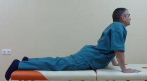
The patient lies on his stomach. At the puncture site, the skin is disinfected and anesthetized with a local anesthetic. Depending on the puncture site, when the needle is inserted, the patient can sit, lie on his side or on his stomach. The needle is advanced inward under the control of a fluoroscope, and after the injection of contrast material is removed, the skin is disinfected again. The patient remains face down on the table.
The doctor slowly tilts the table so that the contrast agent spreads over the subarachnoid space.
To obtain X-ray images, the patient turns on his side and at the time of the image must remain as still as possible to reduce the likelihood of image blur.
Some time after the end of the procedure, you need to wait while the doctor analyzes the received images. In general, myelography takes 30-60 minutes.
The video shows the progress of the procedure:
It doesn't hurt at all!
When the anesthetic is injected, the patient feels a prick; when the needle is inserted into the canal, light pressure.
At the time of administration of a contrast agent, the following sensations may appear:
- short-term burning;
- dizziness, headache;
- feeling of warmth;
- redness of the face;
- salty taste in the mouth.
When the introduction of the substance is over, nausea may appear, less often - vomiting.
After the procedure

After the completion of the procedure, the patient is transferred to the ward and monitored for 2-4 hours. All this time, he should lie with the headboard raised (about 45 °) and observe an increased drinking regime, which will help to quickly remove the contrast agent.
The first 2 days after myelography, intense physical activity and bending are contraindicated. If the temperature rises to 37.5 ° C or more, the appearance of nausea or vomiting, persistent severe headaches, impaired urination and defecation, neck stiffness or numbness of the legs, you should immediately inform the doctor.
results
Myelograms are interpreted by a radiologist or other physician qualified to interpret the results of such examinations. After studying the images, a conclusion is drawn up, which is discussed with the attending physician and is considered taking into account the anamnesis. In some cases, an additional examination is carried out if the results obtained are in doubt and require additional images or the use of special imaging techniques.
Research prices in Moscow
Today in Moscow, myelography is performed in several multidisciplinary medical centers:
- KB MGMU Sechenov (Frunzenskaya, st. B. Pirogovskaya, 2, p. 4);
- JSC "Medicine" (Mayakovskaya, lane 2 Tverskoy-Yamskaya, 10);
- Central Design Bureau No. 1 of JSC "Russian Railways" (Tushinskaya, Volokolamskoe highway, 84);
- Central Design Bureau of Civil Aviation (Tushinskaya, Ivankovskoe highway, 7);
- Central Clinical Hospital FCS (Shchelkovskaya, Otkrytoe shosse, 32),
as well as in many private clinics specializing in such research.
Examination prices vary widely, since they largely depend on the level of the clinic and the material used. Today the cost of myelography is 3000-6000 rubles.
1. Recording
Research is carried out by appointment. You can sign up by phone. Or order a call back
Make an appointment
Note!
The magnet tunnel is a 2 meter long pipe with a diameter of 55 cm. Patients with dimensions exceeding the diameter of the tube cannot be examined with a closed tomograph. You can visit our center prior to your appointment to ensure that size restrictions or claustrophobia will not interfere with your exploration. If you are unable to remain motionless for a long time or suffer from claustrophobia, please report this when recording.
If you are unable to remain motionless for a long time or suffer from claustrophobia, please report this when recording. During the appointment, inform the operator of the list of studies you are interested in, last name, first name, patronymic and date of birth. Fill in the date, time and place of research in the referral form, this will help you avoid surprises and will allow you to visit the center at the time reserved for you.
2. Preparation for research
Most MRI examinations do not require special training, which means that you can eat, drink, and take prescribed medications both before and immediately after the exam. Wear comfortable, metal-free clothing and bring a change of footwear. Leave your jewelry and bijouterie at home. Before the examination, it is not recommended to use cosmetics containing metal particles and metal-containing ointments.
MRI of the abdominal and retroperitoneal organs
- Taking the drug "Espumisan" - to eliminate increased gas formation.
- The study is carried out on an empty stomach: the last meal should take place no later than 6 hours before the start of the study.
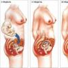
MRI of the pelvic organs in women
- It is recommended to conduct an examination on the 7-12th day of the menstrual cycle, it is possible to conduct a study in the second phase of the cycle with endometriosis and assess the spread of a previously identified tumor process. The study is not carried out during the period of menstruation, it is recommended to calculate in advance the day of the cycle on which the day of the examination will fall.
- For the most qualitative and complete description by the doctor of the results obtained, it is necessary to have with you all the available medical documentation: postoperative extracts, data (images and conclusions) of previous studies of MRI, ultrasound, SCT, and a referral from the attending physician is also desirable.
- The day before the study, coarse fiber and foods that cause excessive gas formation (cabbage, fruits, carbonated drinks, black bread, dairy products, etc.) should be excluded from the diet.
- Taking the drug "Espumisan" - to eliminate increased gas production.
- 30-40 minutes before the start of the study - taking 1-2 tablets "No-shpa".
- Do not urinate 2 hours before the test. Additional fluid intake is not necessary - the bladder should be moderately full
MRI of the pelvic organs in men
- For the most qualitative and complete description by the doctor of the results obtained, it is necessary to have with you all the available medical documentation: postoperative extracts, data (images and conclusions) of previous studies of MRI, ultrasound, SCT, and a referral from the attending physician is also desirable.
- The day before the study, coarse fiber and foods that cause excessive gas formation (cabbage, fruits, carbonated drinks, black bread, dairy products, etc.) should be excluded from the diet.
- Bowel preparation: with normal digestion - natural bowel emptying on the day of the study, with constipation - the use of gentle microclysters (Mikrolax)
- The last meal should be no later than 4 hours before the start of the study.
- 30-40 minutes before the start of the study - taking 1-2 tablets "No-shpa".
- Do not urinate 2 hours before the test. Additional fluid intake is not needed - the bladder should be moderately full.
MRI of the rectum
- Bowel preparation: with normal digestion - natural bowel emptying on the day of the study, with constipation - the use of gentle microclysters (Mikrolax)
- An hour before the study - emptying the bladder and further exclusion of fluid intake.
- 50 minutes before the examination, take 2-3 tablets "No-shpa"
- You must have with you all the medical documentation related to this disease: referral from the attending physician, results of colonoscopy or sigmoidoscopy, postoperative discharge, results of previous MRI or CT examinations
MRI Guided Prostate Biopsy Preparation
- Stop taking anticoagulants 7 days before the biopsy (aspirin, thrombosis, cardiomagnyl, warfarin, Plavix, Clexane, Marevan, Pradaxa, Syncumar, Fraxiparin, Fragmin
and etc.) - Ciprofloxacin 500mg x 2 r / day. - start taking 1 day before the biopsy and then take another 4 days (only 5 days of taking).
- Metronidazole 250mg x 3 r / day - start taking 1 day before the biopsy and then take another 4 days (only 5 days of taking).
- On the evening before the biopsy and in the morning on the day of the biopsy - a cleansing enema.
- 2 hours before biopsy do not urinate.
Myelography makes it possible to monitor the spread of cerebrospinal fluid within the boundaries of the spinal membranes.
With its help, it is possible to identify pathologies of the spinal column, a problem with the nerve roots and spinal cord, and also to clarify the location of the resulting disorders and their causes.
What is this diagnostic method used for?
Usually, during a spinal cord examination, doctors will prescribe. When the subject has a pacemaker, MRI is prohibited. For such cases, myelography is used as a diagnosis of pathological conditions.
Typically, this procedure is done to locate the spinal canal, nerve roots, and blood vessels that supply blood to the spinal cord.
Problems that can be detected with myelography:

It can also be used as an additional procedure for MRI to detect the following conditions:
- tumor spine, lining of the spinal cord, nerve roots and tissues itself;
- infections affecting the spine, problems with the intervertebral membranes of the spinal cord and the tissues that surround it;
- inflamed arachnoid that covers the spinal cord itself;
- pathological changes in the spine and spinal cord subsequently formed from injuries and other diseases.
Myelography also helps during the planning of the operation, as it allows you to predict the effect of the surgical intervention in each individual variant.
Contraindications to diagnosis
There are certain contraindications to this diagnostic method:
- inflammatory or infectious diseases at the puncture site;
- difficulties in the administration of contrast agents;
- the patient is not able to lie still;
- during pregnancy.
You should also inform your doctor about the following conditions:
- , convulsions;
- diabetes;
- kidney disease;
- bronchial asthma;
- problems with blood clotting;
- allergy to drugs.
Pros and cons of the procedure
The advantage of this diagnostic method is that it is one of the most proven and safe methods for human health. Using myelography, the doctor can have images of the smallest spinal vessels, which is almost impossible to see using other methods. The substance introduced into the body is removed quite simply and absolutely does not harm the body.
Among the disadvantages, it can be noted that at the end of the study, there may be frequent headaches, dizziness and severe weakness.
Also, the injected contrast agent can create feelings of nausea, heartburn, vomiting and other problems of the digestive system. Very rarely, the procedure can cause allergic reactions that need to be eliminated by a doctor.
Preparation for research
Instructions for preparation in the procedure should be provided exclusively by the doctor who will perform the myelogram. It is required to tell the doctor about the medications that were taken by the patient, and about the allergy to medications that contain iodine.
The doctor must be informed about the transferred operations and chronic diseases.
Two to three days before the start of the procedure, it is required to stop taking drugs, which include antipsychotic drugs, antidepressants, drugs that thin the blood, and drugs used in the treatment of diabetes mellitus.
The most important thing is to stop taking anticoagulants, which tend to thin the blood. If it is not possible to stop taking medications, then the doctor is required to prescribe other methods of maintaining blood viscosity during the procedure.
Some anticonvulsant drugs must be canceled before the study, as well as warn about the presence of diseases that are accompanied by seizures.
The day before the procedure, you need to increase the amount of fluid you drink, since during this period, sufficient hydration of the body is required. Before the study, you need to stop eating solid food, but you should not limit the liquid.
Change into a hospital gown for the duration of the study. Jewelry, removable dentures and hardware can also affect the resulting image.
Research progress
Myelography is always performed on an outpatient basis. The subject lies on his stomach. To obtain images, a fluoroscope is used, which makes it possible to examine the spine and find the best point for entering contrast.

Contrast input
The material should be injected into the spinal canal just below the lumbar spine, as this is a less dangerous and more convenient option. When the doctor thinks it would be better to use another option, the contrast can be injected at the level of the upper cervical spine.
Where there was a puncture, the doctor must disinfect the site and anesthetize it with a local anesthetic. The subject lies on his side, stomach, or sits down, the choice of posture depends on the localization of the puncture. With the help of a fluoroscope, the doctor moves the needle inward by the time it enters the subarachnoid space.
The hit signal will be the appeared drops of cerebrospinal fluid. The doctor can send this liquid for research in the laboratory. Next, you need to enter the required material through the game, after which it is taken out and the skin is re-disinfected. After that, the subject lies down, most often on his stomach.
Further, the radiologist, using a fluoroscope, slowly changes the inclination of the table, which makes it possible for the introduced material to disperse to the sides along the subarachnoid space, thereby highlighting the outlines of the spinal cord and nerve roots. To remove X-rays, the subject will have to lie on his side. It is important to remain as still as possible at the time of capture, as this reduces the risk of blur.
Myelography is a relatively painful procedure. Sometimes only a headache can last up to several days. Cases of nausea, difficulty breathing, and dizziness are also common. During the examination, a burning sensation may occur along the spine.
 Where there was a puncture, itching, inflammation of the skin lining and even bleeding are possible.
The decoding of the images obtained is carried out by a radiologist who is engaged in X-ray studies and the identification of their results. When the results are examined, the radiologist writes out a report.
The results can be explained by the attending physician, and if the data obtained are doubtful, then an additional examination is required.
Purpose of myelography
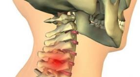
X-ray examination is one of the most important methods in the diagnosis of many diseases, including diseases of the spinal cord and spine. Unfortunately, a simple X-ray of the spine gives little information about the state of the structures of the spinal cord, nerve roots and spaces of the spinal canal, since only bone structures can be seen on a simple X-ray. To improve the results of radiography, radiopaque substances are used. They absorb X-rays and allow you to see certain structures that are not visible on a conventional X-ray.
Myelography is an X-ray contrast method for examining the spine and structures of the spinal cord, in which a radiopaque substance is injected into the spinal canal using a needle. This substance is injected into the subarachnoid space, that is, between the arachnoid and the pia mater, which allows the doctor to see on an X-ray both the structure of the space itself and the nerve roots.
When a radiopaque contrast agent is injected into the subarachnoid space, the doctor can see the spinal cord, nerve roots, and the membranes that surround the spinal cord and nerve roots.
At the same time, the radiologist sees the passage of the radiopaque contrast agent in real time within the subarachnoid space, and can also take pictures. In most cases, computed tomography is done after myelography for even more detailed and layer-by-layer images.
Myelography gives a detailed picture of the condition of the spinal cord and spine.
Indications for myelography
Myelography is most often used to diagnose pathology of the spinal cord, spinal canal, spinal nerve roots and blood vessels that feed the spinal cord:
- To detect the presence of a herniated disc, which compresses the nerve roots or spinal cord.
- To identify spinal stenosis (degenerative changes in the bone and soft tissues around the spinal canal). In this condition, the spinal canal narrows due to an increase in the surrounding bone structures in the form of growth of osteophytes and adjacent ligaments.
Myelography can also be used to assess the following conditions when MRI cannot be used, or in conjunction with MRI:
- Spinal cord tumors.
- Infectious processes.
- Inflammation of the arachnoid membrane that covers the spinal cord.
- Spinal stenosis caused by injury or other medical condition.
Abroad, the very first research method in diagnosing pathology of the spinal cord and spinal column is magnetic resonance imaging (MRI or MRI). This is not always possible, for example, when the patient has built-in medical devices, such as pacemakers, or metal structures in the limbs (wires, plates, bolts, artificial joints), or when an MRI scan is not possible. In some cases, myelography is performed in combination with computed tomography and / or MRI for a more detailed diagnosis.
Myelography can show whether surgical treatment will be effective in this case, and if the answer is yes, it helps to plan surgery.
Preparing for myelography
Your doctor will give you detailed instructions on how to prepare for myelography.
You should inform your doctor about any drugs you are taking, whether you are allergic to drugs or other allergens, especially barium or iodine-containing X-ray contrast materials. Tell your doctor about any previous illness.
Most importantly, your doctor needs to know what drugs you are taking so that you can stop taking them a few days before your myelography procedure and if you have had any previous episodes of allergy to radiopaque contrast agents.
Some medications before myelography should be stopped a couple of days before the procedure. These include some antipsychotic drugs, antidepressants, blood thinners, and diabetes medications, especially metformin. Some of the important drugs on this list are blood thinners (anticoagulants). If you are taking these medications, you should inform your doctor so that alternative medications can be selected while preparing and performing the myelography procedure. For example, after the use of traditionally long-acting anticoagulants has been discontinued, heparin may be used.
Some anticonvulsants should also be discontinued before myelography. This is why it is so important to tell your doctor if you have a seizure or if you are taking any medications for this, so that the doctor can help you stop taking these medications while preparing and performing myelography.
Although an allergic reaction to iodine-containing X-ray contrast agents is very rare, you should still inform your doctor if you have had any previous episodes of allergy to such X-ray contrast agents or other drugs. You should inform if you are allergic to other substances and do not suffer from bronchial asthma. In this case, you will be closely monitored when injecting a radiopaque contrast agent. Allergy to iodine-containing radio-opaque substances is a very dangerous phenomenon.
Usually, patients are advised to drink more fluids before myelography the day before the procedure. Solid food a few hours before myelography is not recommended, but liquids should be continued.
You will be asked to take off your clothes and gown, jewelry, glasses, and other metal objects that might interfere with myelography.
Women should always inform their doctor before undergoing myelography if they are pregnant or expecting to become pregnant. X-ray examinations, including myelography, are not performed during pregnancy to avoid the risk of adverse effects on the fetus.
At the end of the myelography procedure, the patient remains in the ward under observation for 1-2 hours, after which he can be released home.
Equipment used for myelography
For myelography, X-ray equipment, an intravenous infusion system, syringes and needles are used to inject a radiopaque contrast agent into the subarachnoid space. The resulting image can be recorded both on X-ray film and on a digital storage medium, or on both at once.
Principle of myelography
In conventional radiography, X-rays are retained by bone tissue, while all other soft tissues - the spinal cord, its membranes, nerve roots and intervertebral discs - let these rays pass freely. When a contrast agent is injected into the subarachnoid space, it retains X-rays more strongly than bone tissue. Areas where the contrast agent does not enter are allowed to transmit X-rays. As a result, the contours of all soft tissues are obtained - the spinal cord, its membranes, nerve roots.
The resulting image is stored on X-ray film, or, as in most modern clinics, on a digital carrier - disk, flash card.
Myelography method
Usually, myelography is done on an outpatient basis.
The patient lies face down on a special table and the radiologist uses fluoroscopy to determine the best place to inject the contrast media.
The radiopaque contrast agent is injected in the lumbar region, as this is the safest and easiest method to inject. In case of difficulty, the injection of a contrast agent can be carried out in the cervical spine.
At the injection site, the skin is treated with an antiseptic and anesthetized with a local anesthetic. Depending on where the radiopaque contrast agent is to be injected, the patient may lie on his side with his knees pressed to his stomach, or sitting hunched over. In some cases, the patient can sit up. The needle is inserted under fluoroscopy control and advanced until it reaches the subarachnoid space in the spinal canal. At the same time, cerebrospinal fluid begins to slowly drip from the needle. If necessary, a certain amount of this liquid can be taken for analysis. Then a radiopaque substance is injected into the subarachnoid space, after which the needle is removed, and the puncture site is again treated with an antiseptic. Then the patient lies on his stomach on a special table of the X-ray machine.
Then, under the control of the fluoroscope, the radiologist slowly tilts the table so that the radiopaque substance fills the entire subarachnoid space. As the table is tilted, the doctor controls the contrast movement with the fluoroscope, paying particular attention to the area of the alleged lesion. At this point, the patient can be turned to the other side for additional images.
During myelography, it is important that the patient remains motionless so that the images are not blurred. When the imaging is finished, the stage returns to a horizontal position and the patient can turn on his back.
Usually, after myelography, computed tomography is performed immediately, while there is a radiopaque contrast agent in the subarachnoid space. This combination of the two research methods is called CT myelography.
Typically, the myelography procedure takes 30 to 60 minutes. Computed tomography takes an additional 15 - 30 minutes for a complete examination.
Feelings during and after myelography
When a local anesthetic is injected into the skin to numb the puncture site, you will feel a small prick and some bloating sensation and sharp pain when the needle is inserted into the spinal canal.
During the exploration, you will be asked to lie as still and as still as possible while the table tilts at different angles. The legs are usually secured with straps to keep them from moving. While lying face down, you may feel some discomfort, however, be patient, because you will not lie there for long. Less commonly, while the patient is lying face down, difficulty breathing or swallowing may be felt. In this case, you need to inform the radiologist about this, and the table will be raised to a more comfortable position.
Headaches, hot flashes, or nausea may occur after a radiopaque contrast agent is injected, but very rarely. The risk of seizures is also possible, but this is extremely rare with the use of modern X-ray contrast agents.
After the completion of myelography, the patient is transported on a gurney to a special ward, where he is recovered and monitored for one to two hours. In some cases, the patient is left to lie with the raised head end at an angle of 30 ° to 45 ° degrees for four hours. In order for the radiopaque substance to leave the body sooner, you should drink more fluids, this helps to prevent headaches.
After myelography, physical activity and bending of the spine (bending, lifting weights) should be avoided for 1–2 days.
In case of fever and chills, nausea and vomiting, severe headache that lasts more than a day, neck stiffness or numbness in the legs, you should inform your doctor. In case of problems with the administration of physiological needs, you should also consult a doctor.
Processing of myelography results
The results of myelography are interpreted by a radiologist. After a detailed analysis of the images, he sends them with a conclusion to your doctor.
Benefits of myelography
- Myelography is a relatively safe and painless research method.
- When a radiopaque material is injected into the subarachnoid space, it allows the doctor to see all the structures of the spinal cord, including its membranes and nerve roots, which makes it possible to evaluate the different parts of the spine that are not visible on a simple x-ray.
- X-rays at the dose used in myelography are safe for the patient.
Disadvantages of myelography and risk of complications
- X-rays still have, albeit a very low, risk as a carcinogen. However, the benefits of this research far outweigh this risk.
- The effective dose of radiation can vary.
- Although rare, there is a risk of headache after myelography associated with puncture. In this case, the headache occurs after the patient has sat down or got up. The peculiarity of the headache is that it decreases when the patient lies down. Typically, this headache may occur 2 to 3 days after the myelography procedure. For it to pass, you should lie horizontally on your back and drink more fluids, but for more severe pain, you need to take pain relievers. In some situations, some patients continue to have headache, which may require a special but simple procedure for stopping the outpouring of cerebrospinal fluid from the puncture site.
- Adverse reactions to the introduction of a radiopaque contrast agent during myelography are rare and usually mild. They can manifest as a rash, itchy skin, sneezing, nausea, or anxiety. Rarely, urticaria and shortness of breath may appear, which already requires medical intervention.
- Other rare complications of myelography include trauma to the nerve with a puncture needle and bleeding in the area of the nerve roots, where they exit from the spinal canal. There is a risk of inflammation or infection of the spinal membranes. Seizures are an extremely rare complication of myelography. There is a very low risk of a change in pressure in the spinal canal caused by the introduction of a needle below the obstruction site leading to a blockage of cerebrospinal fluid movement in the subarachnoid space, which requires urgent surgical intervention.
- Women should always tell their doctor if they are pregnant or suspect.
Limitations for myelography
- The most significant limitation of myelography is that it allows you to see only what is "going on" inside the spinal canal and in the area of the nerve roots until they leave the canal. The pathology outside the spinal canal is not visible on the myelogram, which is why computed tomography or magnetic resonance imaging is required.
- Myelography is usually not performed in pregnant women due to the possibility of the risk of negative effects on the fetus of x-rays.
- If the patient was unable to lie still during myelography, the images may be blurry.
- In patients with structural defects of the spine after trauma, the introduction of a radiopaque contrast agent into the spinal canal may be difficult.
- Myelography is not performed for infectious and inflammatory skin diseases at the site of the intended injection of the needle.
(495) 50-253-50 - free consultation on clinics and specialists
- Spine myelography