Symptoms and treatment of emphysema. Pulmonary emphysema - symptoms of primary and secondary, mechanism of disease development and signs on x-ray Bullous emphysema x-ray description protocol
Patients with lung diseases are familiar firsthand with the manifestations of respiratory failure. Shortness of breath, decreased exercise tolerance, dry cough and cachexia can be caused by increased pulmonary airiness. What is pulmonary emphysema, why does it develop, how is it diagnosed and treated? You will find answers to these questions in our detailed review and video in this article.
The term “pulmonary emphysema” in medicine combines a number of pathological processes characterized by an increased content of air in the lung tissue. This chronic disease is accompanied by disruption of external respiration processes, as well as gas exchange in the alveoli.
As a result:
- the partitions between the alveoli are destroyed;
- the diameter of the distal branches of the bronchi and bronchioles increases;
- The volume of lung tissue increases, and air voids appear in it.

The lungs literally inflate, stretching the chest along with it, which takes on a characteristic barrel-shaped shape.
This is interesting. The name of the pathology comes from the Greek word “emphysao” - “to inflate”, “to blow”.
Emphysema – ICD 10 code – J43.9 – is a fairly common pathology among the elderly. Along with bronchial asthma and chronic obstructive bronchitis, it is included in the COPD group.
All of them are accompanied by disturbances in air conduction through the respiratory tract, which determines the similarity of their clinical manifestations. However, each of the nosologies has its own distinctive features, which must be taken into account during differential diagnosis.

Causes and pathogenesis
All significant causes of pulmonary emphysema can be divided into two large groups.
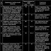
Table 1: Possible reasons emphysema:
| Group | Etiological factor | Description |
| Disorders associated with loss of lung tissue elasticity | Congenital malformations of the respiratory system | Various defects in the formation of bronchioles during the prenatal period can lead to collapse of bronchioles and increased pressure in the alveoli |
| Hereditary alpha-1 antitrypsin deficiency | A decrease in the concentration of the substance causes pathological activation of proteases and destruction of the pulmonary alveoli | |
| Hormonal imbalance | Violation of the physiological relationship between male and female sex hormones leads to stretching of the bronchioles | |
| Living in an environmentally unfavorable region | Tobacco smoke, smog, coal dust, nitrogen and sulfur oxides are deposited on the walls of small bronchi, causing destruction and “bloating” of lung tissue | |
| Frequent lung infections | The development of pneumonia or bronchitis triggers a cascade of immune reactions designed to destroy pathogenic bacteria. By-effect such activity consists in the destruction of the alveolar wall by its own macrophages or neutrophils | |
| Age-related changes | In older people, blood circulation in the lung tissue deteriorates and the regenerative capabilities of the body decrease | |
| Disorders caused by increased pressure in the lungs | Occupational hazards | Increased pressure in the alveoli is created during work at:
|
| Chronic obstructive bronchitis | Thick viscous sputum and broncho-obstruction cause obstruction in the pulmonary tree, as well as overstretching of the bronchioles | |
| Blockage of the bronchial lumen by a foreign body | Prevents air from escaping from a particular segment or lobe of the lung and causes acute emphysema |
In the pathogenesis of the disease, several sequential processes can be distinguished:
- Increased lumen, overstretching of alveoli and bronchioles.
- Thinning of the walls of the vessels supplying the acinus, poor circulation.
- A decrease in the area for gas exchange formation due to the filling of some alveoli with a gas mixture containing a high percentage of CO2.
- Increasing phenomena of local hypoxia.
- Compression of functioning lung tissue by expanded areas, ventilation disturbances.
- Increased intrapulmonary pressure, restructuring of the circulatory system and the formation of chronic pulmonary heart disease.
- Hypoxia of all organs and tissues, signs of respiratory failure.

Simply put, with emphysema, a kind of “stagnation” of air occurs in the lungs. Due to the reasons described, it enters the alveoli, but cannot leave them in the same volume. At later stages, both the process of exhalation and the process of inhalation are disrupted.
Classification
In medicine, it is customary to distinguish several classifications of pathological syndrome.
Depending on the origin it is:
- vicarious (compensatory), which develops after resection of part of the lung;
- senile, developing against the background of age-related involution of the body;
- primary, which is rare and is accompanied by severe atrophy of the alveolar wall;
- interstitial, occurring when the alveoli rupture (for example, with a strong cough).
According to prevalence, pathology is divided into diffuse and focal. In the focal form, changes in the pulmonary parenchyma occur around pathological foci of tuberculosis and cicatricial changes.
Widespread emphysema is characterized by the destruction of alveoli throughout the lung surface and is associated with severe chronic diseases. Diffuse emphysema in children appears due to obstruction of one of the large bronchi.
Depending on the anatomical features, the following pathology development options are distinguished:
- armored– is severe, characterized by widespread lesions and the absence of healthy lung tissue between the swollen acini;
- centrilobular, in which destructive processes affect only the central area of the acinus;
- panlobular emphysema characterized by uniform damage to the pulmonary lobule and more extensive areas of destruction;
- periaciar(more common in tuberculosis) – accompanied by damage to mainly the distal parts of the acinus near the pleura;
- bullous– characterized by the formation of bullae – large bubbles, the diameter of which can reach 5-200 mm.
- interstitial, in which the alveoli rupture and air accumulates in the tissues of the interstitium (interlobular tissue along the bronchi);
- subpleural, accompanied by the accumulation of air under the pleura;
- interstitial (subcutaneous), characterized by the accumulation of released air under the skin.

Symptoms: how does increased airiness of the lung tissue manifest?
Depending on the form and extent of the lesion, the symptoms of the pathology may have different severity.
Common signs of the disease include:
- shortness of breath;
- sometimes – cough with light mucous sputum;
- barrel chest;
- decrease in the amplitude of movement of the HA during breathing;
- weakening of breathing;
- manifestations of respiratory failure:
- expansion of intercostal spaces;
- bulging of the supraclavicular fossa;
- cyanosis;
- swelling of the neck veins;
- puffiness of the face.
Note! In patients with primary emphysema, one can often observe the “puffing” syndrome - covering the mouth while exhaling with swelling of the cheeks.
The primary increase in airiness in the alveoli is always more severe than the secondary one. It is accompanied by severe shortness of breath and severe discomfort for the patient. However, the gas composition of the blood in secondary forms of the disease has more pronounced disturbances.
The progressive course of pulmonary emphysema can lead to complications such as:
- respiratory failure;
- pulmonary hypertension;
- heart failure(mainly due to dilatation of the right ventricle): hepatomegaly, ascites, edema.

Important! Bullous emphysema can cause repeated episodes of spontaneous pneumothorax.
Diagnostic principles
Diagnosis of emphysema requires integrated approach. If the patient has complaints or clinical manifestations of the disease, he should be examined by a doctor: a therapist or a pulmonologist.
Standard medical instructions imply the following diagnostic algorithm:
- Collection of complaints and medical history. At this stage, it is important for the specialist to find out whether the patient has shortness of breath and cough, and how long they last.
- Percussion. Using a special technique of tapping the chest with your fingers, the doctor can determine:
- limited mobility of lung tissue;
- “boxy” sound when percussing over areas of increased airiness;
- reducing the boundaries of relative dullness of the heart;
- prolapse of the lower pole of the lungs.
- Auscultation with a medical stethoscope. Revealed:
- Increased exhalation;
- Tachypnea – increases
- Muffling of heart sounds;
- Decreased breathing;
- Sometimes – dry wheezing over the entire pulmonary surface;
- Compensatory tachycardia.
- Instrumental studies:
- Radiography;
- Scintigraphy;
- Spirometry;
- Peak flowmetry;
- Study of blood gas composition.
Note! If it is difficult to make a diagnosis based on the results of R-graphy, the patient may be prescribed more modern look examinations. On a CT or MRI, the images are clearer, and it is also possible to take a layer-by-layer image of the lungs.
Features of treatment
Among the main goals of treatment of emphysema are:
- combating the underlying pulmonary disease;
- prevention of complications;
- improving the patient's quality of life.
Mandatory treatment measures for all patients with increased airiness of the alveoli include treatment of the underlying disease, prevention of respiratory system infections and smoking cessation. The therapeutic diet for pulmonary emphysema involves split meals in small portions.
Useful products are considered:
- all kinds of cereals;
- unprocessed seasonal vegetables and fruits - carrots, zucchini, pumpkin, broccoli, all citrus fruits, etc.;
- dried fruits (figs, prunes, dried apricots);
- seafood;
- sources of quality protein - lean meat, fish, dairy products, legumes;
- herbal teas and decoctions of hawthorn, rose hip, and linden leaves.

Regular exercise is also useful therapeutic exercises, which will improve lung ventilation and reduce hypoxia.
Table 2: Exercise therapy:
Note! This series of exercises should be performed 3-4 times a day.
In addition, walking, swimming, and cycling can reduce the manifestations of respiratory failure and saturate the body with the necessary oxygen. At the same time, during physical activity You should breathe through your nose, not your mouth.
Medications
Plan drug therapy compiled by a doctor individually for each patient. The main groups of drugs used in treatment are presented in the table below.
Table 3: Main medications for emphysema:
| Pharmacological group | Mechanism of action | Representatives |
| α1-antitrypsin inhibitors | Reducing the number of proteases that destroy pulmonary alveoli | Prolastin |
| Mucolytics |
|
Alethylcysteine |
| Ambroxol | ||
| Antioxidants |
|
Tocopherol |
| Phosphodiesterase inhibitors |
|
Teopek |
| Anticholinergic drugs |
|
Atrovent |
| Theophyllines |
|
Theophilus |
| GCS (according to indications) |
|
Prednisolone |

Inhalations
Inhalations also have important. To eliminate hypoxia, breathing through an oxygen mask is indicated. For maximum effect, it is important to continue the procedure for at least 18 hours in a row.
Concomitant chronic inflammation requires inhalation with a nebulizer: with emphysema in combination with bronchitis, you can breathe a solution of Ambroxol, Belodual or other bronchodilators.
When surgery may be required
Surgical treatment of emphysema is rarely performed, only if drug therapy is ineffective.
The main indications for surgery are:
- multiple bullae occupying more than 1/3 of the lung area;
- shortness of breath that cannot be controlled with medications;
- development of acute complications of the disease: pneumothorax, hemoptysis, malignant neoplasm;
- the need for frequent hospitalizations;
- progressive deterioration of the condition.

Among the surgical techniques used in the treatment of emphysema, the most common are:
- lung transplantation;
- removal of part of the lung tissue through thoracoscopy or abdominal surgery.
As a result of surgical intervention, lung ventilation is restored, shortness of breath is reduced, and tolerance to physical activity significantly increases.
Important! Severe cachexia, concomitant diseases in the stage of decompensation, and old age may be considered contraindications for surgery.
According to statistics, emphysema and pulmonary pneumosclerosis are the most common diseases that cause chronic respiratory failure. Moreover, the pathological changes that occur with them are mostly irreversible. Therefore, all that remains for the pulmonologist is to slow down their progression, reduce the severity of DN and alleviate the patient’s condition.
The prognosis for pulmonary emphysema largely depends on:
- timely diagnosis and adequate treatment of the underlying disease;
- an integrated approach to the treatment of hyperairy lung syndrome and its complications;
- compliance by the patient with all prescribed recommendations;
- features of the course and duration of the disease.
Usually, the manifestations of emphysema are easily corrected if the patient refuses bad habits(in particular smoking), timely prevention of complications and pulmonary infections. Living in a clean air environment is also important for the success of therapy.
Questions for the doctor
Causes of shortness of breath
My dad is 78 years old. He complains of severe shortness of breath, which prevents him from moving around the house calmly. We took dad to the clinic and did a cardiogram - everything was normal. The therapist suggested that he had emphysema and sent him for examination (X-rays, tests). What kind of disease is this? Isn't this cancer?
Hello! Pulmonary emphysema is a serious condition accompanied by increased airiness of the alveoli and their subsequent destruction. It is most often diagnosed in older men.
It has many various reasons. These include genetic defects, long-term smoking, and chronic bronchitis. The statement that emphysema is lung cancer is incorrect, but sometimes these serious diseases are diagnosed in patients together.
Therefore put accurate diagnosis your dad and the cause of shortness of breath can only be determined after a comprehensive diagnosis.
Disease prevention
My friend has been coughing for several months now, and Lately severe shortness of breath began to bother me. I went to the doctors, was examined, and was diagnosed with emphysema. He explained to me that this was due to the accumulation of air in the lungs. But I'm worried. Is emphysema contagious? And how to protect yourself from this disease?
Hello! No, emphysema is not an infectious disease, so the risk of catching it from a friend is zero. Prevention of the disease consists of quitting smoking, timely treatment of respiratory tract infections, maintaining a healthy lifestyle, and walking in the fresh air.
Chronic obstructive pulmonary disease (COPD) is a chronic pathology caused by damage to the distal respiratory tract and pulmonary parenchyma with the development of emphysema, manifested by partially reversible bronchial obstruction. The following factors contribute to the development of COPD: genetic predisposition, respiratory infectious diseases, smoking, influence of inorganic and organic dust.
Inflammation in COPD causes partially reversible bronchial obstruction. The irreversible component of bronchial obstruction is caused by the development of fibrosis in the bronchial wall. Against the background of this process, the development of pulmonary emphysema occurs, characterized by the destruction of the alveolar walls and the formation of bullae (thin-walled cavities) in the lungs. Infectious diseases of the respiratory tract play an important role in the development of COPD.
With COPD, the patient develops shortness of breath, cough with sputum, and dry wheezing is heard in the lungs on auscultation. Severe pulmonary emphysema is manifested by an increase in the anteroposterior volume of the chest (the so-called “barrel” chest). Also, to diagnose the condition, a respiratory function test is used, which determines obstructive disorders in COPD.
COPD is characterized by bronchial obstruction, which results in excessive accumulation of air. On a radiograph this is defined as hyperairiness of the lungs. X-ray manifestations of hyperairy lungs have the following signs (Figure 1):
- Sealing and low diaphragm domes
- Increase in the area of the retrosternal space
- "Drip" shape of heart shadow

Figure 1. Chronic obstructive pulmonary disease (COPD). The x-ray shows signs of hyperairy lungs: the location of the diaphragm is noted at the level of the anterior segmentsVII-VIII ribs, diaphragm domes are compacted; an increase in the retrosternal space is determined (see arrows), a “drip” shadow of the heart. There is an enlargement of both pulmonary arteries and their hilar branches - a characteristic sign of pulmonary hypertension
A low location of the diaphragm can be assumed if the peak (highest) point of its dome is visualized at the level of the anterior segment of the 7th rib and below (especially if the radiography was performed in a sitting position). COPD is characterized by a bilateral low position of the diaphragm. Note that the assessment of the location of the diaphragm must be carried out with caution, since in athletes or people with tall asthenic physiques, a low location of the domes of the diaphragm is considered normal.
In COPD, an X-ray taken in a lateral projection is characterized by an increase in the retrosternal space, due to the fact that hyper-airy lung tissue pushes the heart and blood vessels back, while an increase in the anteroposterior volume of the chest is noted ("barrel" chest). A narrow, vertically elongated shadow of the heart (“drip” heart) is determined.
Main feature emphysema - bullae(thin-walled cavities in the lungs, more than 1 centimeter in size (Figure 2).

Figure 2. COPD. A B- X-ray in the right lateral projection. In the lower lobe of the right lung there is a large bulla - a thin-walled cavity (see indicators). X-ray B shows a backward displacement of the vessels of the lower lobe by the bulla, while the pulmonary pattern is not visible against the background of the cavity. The image shows signs of hyperairy lungs: compaction and low location of the domes of the diaphragm, an increase in the anteroposterior volume of the chest. The expansion of the roots due to pulmonary hypertension is determined. In the ligular segments on the left there is a decrease in transparency due to pneumonia
A typical place for the development of bullae is the upper lobes of the lungs. Bullae can reach large sizes and occupy a significant volume of the lung lobe. In some cases, the wall of the bulla cannot be clearly identified on an x-ray and its presence can only be suspected if the pulmonary pattern is absent or significantly depleted (see article) in a limited area of the lung (Figure 3).

Figure 3. Bullous emphysema in very severe COPD. A- radiograph in direct projection; B- X-ray in the right lateral projection. Signs of hyperairy lungs are determined - compaction and low location of the domes of the diaphragm, a significant increase in the area of the retrosternal space, unification of the pulmonary pattern at the periphery of the lungs. In the upper lobe of the left lung, an area devoid of a pulmonary pattern is identified (see arrows); in the lower part on the left, against the background of fibrous changes, an area devoid of a pulmonary pattern is noted (see pointers) - these changes are caused by bullae
The wall of the bulla may rupture, resulting in pneumothorax. With pulmonary emphysema, diffuse depletion of the pulmonary pattern may occur (especially in the periphery of the lungs), but this sign is not reliable, since the picture of the pulmonary pattern greatly depends on the technical conditions of the radiograph and the patient’s inhalation depth. For the diagnosis of bullous pulmonary emphysema, the diagnostic method of choice is X-ray computed tomography (XCT).
Chronic obstructive pulmonary disease is characterized by increased pressure in the pulmonary artery system, resulting in the development pulmonary hypertension, characterized by the expansion of the roots of the lungs due to the pulmonary arteries (the structure of the roots is preserved, their contours are smooth and clear - see Figure 1, 2). Also in the periphery, the caliber of the arteries sharply decreases, and the hilar pulmonary arteries expand - "gauge jump" symptom.
The X-ray picture of COPD may also include changes in the pattern in the hilar regions of the lungs and thickening of the walls of the bronchi. On a radiograph, the bronchi in longitudinal section are defined as parallel stripes ( "tram rails" symptom). The bronchi in the orthoprojection (in cross section) on the radiograph are defined as small ring-shaped shadows. Note that normally, a radiograph can reveal single parallel and ring-shaped shadows in the root zones, caused by the bronchi, while the thickness of their walls does not exceed 1 mm. In case of development of the inflammatory process, the thickness of the walls of the bronchi is 2-3 mm, the internal lumen of the bronchi narrows, the contours of the bronchi are unclear; the contours of the vessels also lose clarity. These changes are called peribronchovascular “couplings”.
Thus, if in COPD an X-ray shows an increase and deformation of the pulmonary pattern with the formation of reticular (mesh) shadows and the formation of peribronchovascular “couplings” in the hilar regions, this may be a sign of both inflammatory changes during an exacerbation of the process and pneumosclerosis.
02.02.2015
Pulmonary emphysema is a chronic nonspecific lung disease, which is based on persistent, irreversible expansion of the air spaces and increased swelling of the lung tissue distal to the terminal bronchioles. Emphysema is manifested by expiratory shortness of breath, cough with a small amount of mucous sputum, signs of respiratory failure, and recurrent spontaneous pneumothorax.
Pulmonary emphysema (from the Greek emphysema - bloating) is a pathological change in lung tissue, characterized by increased airiness due to expansion of the alveoli and destruction of the alveolar walls. Pulmonary emphysema is detected in 4% of patients, and it occurs 2 times more often in men than in women. The risk of developing emphysema is higher in patients with chronic obstructive pulmonary disease, especially after 60 years of age. The clinical and social significance of pulmonary emphysema in pulmonology is determined by the high percentage of development of cardiopulmonary complications, disability, disability of patients and increasing mortality.
Causes and mechanism of development of pulmonary emphysema
Any causes leading to chronic inflammation of the alveoli stimulate the development of emphysematous changes. The likelihood of developing emphysema increases if the following factors are present:
- congenital deficiency of α-1 antitrypsin, leading to destruction of alveolar lung tissue by proteolytic enzymes;
- inhalation of tobacco smoke, toxic substances and pollutants;
- microcirculation disorders in lung tissues;
- bronchial asthma and chronic obstructive pulmonary diseases;
- inflammatory processes in the respiratory bronchi and alveoli;
- features of professional activity associated with a constant increase in air pressure in the bronchi and alveolar tissue.
Under the influence of these factors, damage to the elastic tissue of the lungs occurs, reduction and loss of its ability to fill and collapse. Air-filled lungs lead to sticking of small bronchi during exhalation and obstructive pulmonary ventilation disorders. The formation of the valve mechanism in pulmonary emphysema causes swelling and overstretching of lung tissue and the formation of air cysts - bullae. Ruptures of bullae may cause episodes of recurrent spontaneous pneumothorax.
Emphysema is accompanied by a significant increase in lung size, which macroscopically becomes similar to a large-porous sponge. When examining emphysematous lung tissue under a microscope, destruction of the alveolar septa is observed.
Classification of pulmonary emphysema
Pulmonary emphysema is divided into primary or congenital, developing as an independent pathology, and secondary, occurring against the background of other lung diseases (usually bronchitis with obstructive syndrome).
Based on the degree of prevalence in the lung tissue, localized and diffuse forms of pulmonary emphysema are distinguished.
Based on the degree of involvement of the acinus (the structural and functional unit of the lungs that ensures gas exchange and consists of the branching of the terminal bronchiole with the alveolar ducts, alveolar sacs and alveoli) in the pathological process, the following types of pulmonary emphysema are distinguished:
- panlobular (panacinar) - with damage to the entire acini;
- centrilobular (centriacinar) - with damage to the respiratory alveoli in the central part of the acinus;
- perilobular (periacinar) - with damage to the distal part of the acinus;
- peri-scar (irregular or uneven);
- bullous (if bullae are present).
Particularly distinguished are congenital lobar (lobar) pulmonary emphysema and McLeod syndrome - emphysema of unknown etiology, affecting one lung.
Symptoms of emphysema
The leading symptom of pulmonary emphysema is expiratory shortness of breath with difficulty exhaling air. Dyspnea is progressive, occurring first during exertion and then at rest, and depends on the degree of respiratory failure. Patients with pulmonary emphysema exhale through closed lips while puffing out their cheeks (as if “puffing”). Shortness of breath is accompanied by a cough with the production of scanty mucous sputum. A pronounced degree of respiratory failure is indicated by cyanosis, puffiness of the face, and swelling of the veins of the neck.
Patients with pulmonary emphysema lose significant weight and have a cachectic appearance. Loss of body weight during pulmonary emphysema is explained by high energy costs spent on intensive work of the respiratory muscles. In the bullous form of pulmonary emphysema, repeated episodes of spontaneous pneumothorax occur.
Complications of emphysema
The progressive course of pulmonary emphysema leads to the development of irreversible pathophysiological changes in the cardiopulmonary system. The collapse of small bronchioles during exhalation leads to obstructive pulmonary ventilation disorders. Destruction of the alveoli causes a decrease in the functional pulmonary surface and the phenomenon of severe respiratory failure.
Reduction of the capillary network in the lungs entails the development of pulmonary hypertension and an increase in the load on the right side of the heart. With increasing right ventricular failure, edema occurs lower limbs, ascites, hepatomegaly. An emergency condition for pulmonary emphysema is the development of spontaneous pneumothorax, requiring drainage of the pleural cavity and aspiration of air.
Diagnosis of emphysema
The history of patients with pulmonary emphysema includes a long history of smoking, occupational hazards, and chronic or hereditary lung diseases.
When examining patients with pulmonary emphysema, attention is drawn to an enlarged, barrel-shaped (cylindrical) chest, widened intercostal spaces and epigastric angle (obtuse), protrusion of the supraclavicular fossa, shallow breathing with the participation of auxiliary respiratory muscles. Percussion is determined by the displacement of the lower borders of the lungs by 1-2 ribs downwards, a box sound over the entire surface of the chest. On auscultation, with pulmonary emphysema, weakened vesicular (“cotton”) breathing and muffled heart sounds are heard. In the blood with severe respiratory failure, erythrocytosis and increased hemoglobin are detected.
At X-ray of the lungs an increase in the transparency of the pulmonary fields, a depleted vascular pattern, limited mobility of the dome of the diaphragm and its low location (in the front below the level of the VI rib), an almost horizontal position of the ribs, a narrowing of the cardiac shadow, and an expansion of the retrosternal space are determined. By using CT lungs The presence and location of bullae in bullous emphysema is clarified.
In case of pulmonary emphysema, the study of the function of external respiration is highly informative: spirometry, peak flowmetry, etc. In the early stages of the development of pulmonary emphysema, obstruction of the distal segments of the respiratory tract is detected. The bronchodilator inhaler test demonstrates the irreversibility of obstruction characteristic of emphysema. Also, with FVD, a decrease in vital capacity and Tiffno test is determined.
Blood gas analysis reveals hypoxemia and hypercapnia, clinical analysis reveals polycythemia (increased Hb, red blood cells, blood viscosity). The examination plan must include an analysis for α -1-trypsin inhibitor.
Treatment of emphysema
There is no specific treatment for emphysema. The primary thing is to eliminate the factor predisposing to emphysema (smoking, inhaling gases, toxic substances, treatment of chronic respiratory diseases).
Drug therapy for pulmonary emphysema is symptomatic. Lifelong use of inhaled and tablet bronchodilators (salbutamol, Berotek, Teopek, etc.) and glucocorticoids (budesonide, prednisolone) is indicated. For cardiac and respiratory failure, oxygen therapy is carried out and diuretics are prescribed. The complex of treatment for pulmonary emphysema includes breathing exercises.
Surgical treatment of pulmonary emphysema involves an operation to reduce lung volume (thoracoscopic bullectomy). The essence of the method comes down to resection of peripheral sections of lung tissue, which causes “decompression” of the rest of the lung. Observations of patients after bullectomy show an improvement in lung functional parameters. Lung transplantation is indicated for patients with pulmonary emphysema.
Prognosis for emphysema
The lack of adequate treatment for pulmonary emphysema leads to progression of the disease, disability and early disability due to the development of respiratory and heart failure.
Despite the fact that irreversible processes occur with pulmonary emphysema, the quality of life of patients can be improved by constantly using inhaled drugs. Surgical treatment of bullous pulmonary emphysema somewhat stabilizes the process and relieves patients from recurrent spontaneous pneumothorax.
Prevention of emphysema
An essential aspect of the prevention of pulmonary emphysema is anti-tobacco propaganda aimed at preventing and combating smoking.Tags:
Start of activity (date): 02/02/2015 21:35:00
Created by (ID): 645
Keywords: lungs, bronchi, shortness of breath
On an X-ray of the chest organs (CH) in frontal and lateral projections, with an increase in the airiness of the lungs, the following radiological syndromes are observed:
- enlightenment;
- expansion of intercostal spaces;
- barrel chest;
- deformation of the pulmonary pattern;
- decreased structure of the roots of the lungs;
- smoothness of the contours of the diaphragm domes;
- drip heart.
Attention! The barrel-shaped chest with emphysema is clearly visible in the lateral projection image, which visualizes an increase in the anteroposterior size (the distance between the sternum and the spine).
X-ray of the lungs in a lateral projection: an increase in the anteroposterior size with emphysema is clearly visualized
X-ray morphological symptoms are secondary. They appear due to the expansion of the chest due to an increase in lung capacity.
Other radiomorphological symptoms of excess air in the lung tissue:
- forward deviation of the sternum;
- horizontal arrangement of ribs;
- expansion of the anterior mediastinum;
- symmetrical protrusion of the chest anteriorly.
Radiological symptoms of emphysema are also observed in the lungs:
- Increase in the area of the lung fields.
- Diffuse transparency enhancement.
- Local areas of clearing in places of accumulation of emphysematous bullae.
- Redundancy of the pulmonary pattern.
During the disease, the dome of the diaphragm deviates downwards due to the pressure on it from the increased size of the lungs. With severe disease, the dome of the diaphragm becomes like a “tent” - a pointed roof, with which the shadow of the heart merges.
Functional X-ray diagnostic syndromes
Functional X-ray diagnostic syndromes arise due to increased ventilation in the lung tissue. As the elasticity of the alveoli decreases, their volume increases. As a result, the internal cavity of the alveolar acini is filled with air. X-rays, passing through such anatomical formations, are not delayed, so a clearing is formed in the image.
The difference in X-ray contrast is clearly visible in the lower (basal) parts of the lungs, where active ventilation occurs.
To correctly read a picture of emphysema, radiologists perform the following tests:
- When exposing the patient's chest, the right dome of the diaphragm is covered with a screen so that its upper edge is in the lower part of the rectangle. With emphysema, there is a restriction in the mobility of the diaphragm in a rectangle measuring 5x5 cm.
- Sokolov's method: a series of photographs are taken on a small film (13x18 cm) in different phases of breathing (inhalation, exhalation and while holding the breath). In a healthy person, there is a difference in contrast between these images. With emphysematous lesions of the lung tissue, the difference is not noticeable.
- The method of targeted images involves taking a series of targeted radiographs in areas of pronounced airiness during maximum inhalation, exhalation and respiratory pause.
Sight image of the right half of the chest with emphysema. It shows a total increase in transparency (clarification)
What does an x-ray tell you about increased transparency?
X-rays provide the clinician with a lot of information about the state of the lung fields. Classic plain radiography of the lungs makes it possible to establish a diagnosis, but it does not always correctly reflect the nature of the pathological process that formed the airiness of the pulmonary fields. In such a situation, a non-standard OGK x-ray is used, and computed tomography is used. It is more informative, but is characterized by increased radiation exposure, so it is used only in cases of extreme necessity.
Maximum benefit from computed tomography in cases of suspected bullous emphysema (with the formation of large air cavities). To identify the characteristics of the course of other forms of pathology, it is better to use magnetic resonance imaging.
X-rays can also be used to differentiate between the following types of emphysema:
The primary form is not associated with narrowing of the bronchi. Detecting it early can prevent complications, so radiologists must be very careful when reading images of the lungs.
In the secondary form of the disease, x-rays are less informative, since during the study it is impossible to view the internal structure of the bronchi, where chronic inflammatory changes accumulate.
The localized type of the disease is even more difficult to diagnose. Local small foci of increased airiness are very difficult to identify on images, since the affected areas are small, and X-rays are not reflected from the airy tissue.
No matter how informative an x-ray is when diagnosing emphysema, you cannot rely only on its signs, since the layer-by-layer image is quite deceptive.
CT and X-ray diagnostics of pulmonary emphysema
Signs of emphysema on x-rays
When interpreting radiographs, it is often necessary to differentiate pulmonary emphysema from clearing caused by other reasons. Thus, a dark (in negative photographs) image may indicate not emphysema, but an overexposure. Thus, an incorrect choice of voltage (kV) and current (mA*s) can lead to the fact that the pulmonary fields will be too dark, and the pulmonary pattern will not be visualized (especially in the periphery). In some cases, on the images one lung field appears darker than the other - this situation may be associated with the incorrect position of the screening grid; changes on the radiographs should not be regarded as emphysema.
A “dark” image resulting from overexposure should not be confused with emphysema!
On the left, the difference in the transparency of the lung fields is due to the incorrect position of the screening grid, while on the right, in the lower parts of the lung field, the increased transparency of the lungs on the x-ray is due to emphysema
When performing chest x-rays in patients with a history of mastectomy (on one side), a similar picture can be found - one lung field is darker than the other. IN in this case the situation is associated with an uneven decrease in the volume of tissue through which X-ray radiation passes. If the patient is positioned incorrectly (during rotation) or with scoliosis, an asymmetry in the transparency of the pulmonary fields can also be detected, which is again associated with different volumes of tissue on the right and left sides.
Signs of pulmonary emphysema on radiographs include: “rarefaction” of the pulmonary (vascular and reticular) pattern; sign of “break” of pulmonary vessels (dilated branches of the pulmonary arteries abruptly “break” at the periphery of the pulmonary fields); flattening of the domes of the diaphragm, as well as obliteration of the anterior costophrenic sinus (can be detected on a radiograph in a lateral projection).
Changes in radiographs with pulmonary emphysema can be conditionally divided into 4 types, none of which, however, is pathognomonic, but all of them, together with clinical data, quite accurately make it possible to establish the diagnosis of emphysema. These include: - Changes in the chest. Often with pulmonary emphysema, x-rays can reveal an expansion of the chest, an increase in its anteroposterior and bilateral dimensions. The chest is barrel-shaped, the intercostal spaces are expanded, the posterior segments of the ribs have a horizontal course.
Changes in the lung tissue. The main sign of emphysema on radiographs is an increase in the transparency of the lung fields, which, however, must be critically assessed (the conditions listed above must be taken into account). The pulmonary pattern can be “strengthened” (due to pneumosclerosis) or “sparse” (if there are no signs of pneumosclerosis yet). The root of the lung looks expanded (due to the pulmonary arteries) and has a “comma” shape. When bronchography with pulmonary emphysema, the picture resembles a “tree without leaves” due to the lack of filling of small-caliber bronchi with contrast. Functional radiography reveals no difference in the size of the retrosternal and retrocardial space.
Changes from the diaphragm. With emphysema, the right and left domes of the diaphragm are flattened and deformed (changes in the contour of the shadow of the diaphragm in the form of “folds” and all kinds of bulges can be detected). More often, the anteromedial portion of the dome of the diaphragm on the right side bulges, which creates a “double contour” effect on x-rays.
Changes in the heart and blood vessels (aorta, pulmonary artery). With pulmonary emphysema, one can observe the development of the so-called. “pulmonary” heart, with expansion of the shadow of the right ventricle (shift of the shadow of the heart on radiographs to the right). The arch of the pulmonary artery may also bulge.
Signs of emphysema on computed tomography
Emphysema is usually called dilatation of the airways located peripherally - distal to the terminal bronchioles, with destruction of the walls of the alveoli. There are three main types of emphysema (which can be detected on CT): panlobular (all alveoli of the pulmonary lobe are dilated), centrilobular (only the respiratory bronchioles are dilated), and paraseptal (characterized by distribution along the pleura).
Thus, the main signs of emphysema on computed tomography are: zones of increased pneumatization of lung tissue, which alternate with areas of normal structure and density, having clear contours. The density of these areas during exhalation increases minimally or does not increase at all (compared to normal lung tissue). The diameter of the pulmonary vessels near areas of emphysema is reduced.
The centrilobular type of emphysema is characterized by damage to the respiratory bronchioles, while the peripheral parts of the pulmonary lobule remain unaffected. Thus, with centrilobular pulmonary emphysema, CT scan can detect small zones of increased airiness localized inside normal lung tissue, as well as a decrease in the diameter of the pulmonary vessels. This type of emphysema is often found in smokers.
Centrilobular and panlobular emphysema. On the left, number 1 indicates the dilated terminal bronchiole, number 2 indicates normal alveoli; on the right, the number 3 indicates dilated alveoli
With panlobular pulmonary emphysema, computed tomography reveals dilated alveoli and alveolar ducts. Damage to lung tissue is usually diffuse: areas of swelling merge with each other, normal lung tissue is represented by separate “islands,” and there is a significant decrease in the vascularization of lung tissue. The lower lobes of the lungs are predominantly affected. In the final stage, it becomes impossible to distinguish between centrilobular and panlobular emphysema.
Paraseptal emphysema is characterized by expansion of the alveoli, which are directly adjacent to the pleura, as well as to the vascular-bronchial bundles. Clinically, this type of emphysema is the most favorable, since the volume of damage to lung tissue is small, and lung function is extremely slightly impaired. Most often, the paraseptal type of pulmonary emphysema can be detected by CT scan in the area of the apexes of the lungs, in the area of the costophrenic sinuses, as well as along the course of large vessels and bronchi.
1 – bullous elements are adjacent to the pleura, 2 – located along the bronchi and vessels
Congenital lobar emphysema appears on CT scan of the lungs as a zone with increased pneumatization of the lung tissue and reduced vascularization. Mediastinal shift may also be detected. The cause of lobar emphysema is stenosis of one of the lobar bronchi (most often the left upper lobe, less often the right middle lobe and right lower lobe bronchus). Swire-James syndrome manifests itself on computed tomography as follows: one lung has increased pneumatization and is depleted of blood vessels compared to the opposite side. Swire-James syndrome is the result of damage to the lung in the first years of life as a result of long-term obliterating bronchiolitis.
Schematic representation of areas of increased airiness during computed tomography of the lungs: 1 - pneumatocele - a cavity in the lung (most often of a traumatic nature), which has very thin walls, 2 - a cyst in the lung, has walls of uneven thickness (from 1 to 3 mm on average), 3 – bullae (cavities in the lung with thin walls, resulting from the destruction of the walls of the alveoli), 4, 5 – cavities in the lungs with abscesses, tumors with decay, tuberculosis cavities (have walls of uneven thickness, often irregular shape), 6 – changes in type “honeycomb lung” (areas of bullous and cystic transformation of lung tissue alternate with areas of pneumosclerosis)
Signs of pulmonary emphysema on computed tomography – in the area of the apex of the right lung, areas with increased airiness are visualized, with walls of uneven thickness (pulmonary cysts)
CT signs of emphysema of the apices of both lungs
The patient has signs of pulmonary emphysema. CT. On the right, in the basal sections, multiple areas of increased airiness of the lung tissue are visualized (multiple bullae merging with each other), on the left - multiple bronchiectasis (marked with blue arrows)
Severe pulmonary emphysema. CT. Note multiple bullous elements in the lung parenchyma on both sides, as well as a giant bulla in the left lung
Bullous transformation of the lungs in a patient with emphysema. CT.
Second opinion of medical experts
Send your research data and receive qualified assistance from our specialists!
© Second opinion of medical experts
Emphysema
X-ray diagnostics. X-ray semiotics of pulmonary emphysema is very diverse and reflects certain pathomorphological and pathophysiological changes various stages of this disease. Currently, most authors divide all radiological symptoms of pulmonary emphysema into morphological and functional.
X-ray morphological symptoms, reflecting changes in the shape and size of the difficult cell, are secondary and, as a rule, indicate advanced phases of the course of pulmonary emphysema. The most characteristic sign of severe pulmonary emphysema is the so-called barrel-shaped deformation of the chest (Fig. 3), especially clearly identified when examined in the lateral projection due to the predominant increase in the anteroposterior size, i.e., the distance between the sternum and the spine. Three factors contribute to this: a more horizontal position of the ribs than normal, kyphosis of the thoracic spine and anterior protrusion of the sternum. Protrusion of the sternum - a frequent and important symptom of pulmonary emphysema - is usually combined with another significant symptom - expansion of the anterior mediastinum and its increased transparency (“gaping” of the anterior mediastinum). At the same time, in the lateral projection, a noticeable increase in the distance between the sternum, on the one hand, and the shadow of the heart and great vessels, on the other, is noted. This occurs as a result of the heart and great vessels being pushed posteriorly by the expanded anterior sections of the lungs.
In the anterior projection, a noticeable symmetrical protrusion of the lower parts of the chest can be observed, over which, in severe cases of pulmonary emphysema, a kind of “waist” is formed, as a result of which the chest takes the shape of a bell or an hourglass (Fig. 4).
X-ray morphological symptoms are also observed in the lungs. Along with a general increase in the area of the lung fields (mainly due to expansion of the vertical dimensions) and a diffuse increase in their transparency, local areas of increased transparency may be detected due to the formation of multiple large emphysematous bullae, local emphysema or acute swelling of individual areas of the lung (lobe, segment). These local areas of clearing, observed more often in the basal parts of the pulmonary fields, are of great diagnostic importance.
Most authors consider changes in the pulmonary pattern to be characteristic of pulmonary emphysema - its redundancy, sometimes deformation, since pulmonary emphysema is usually combined with pneumosclerotic changes. Some authors consider the redundancy of the pulmonary pattern to be a consequence of peribronchial and perivascular pneumosclerosis, others - the result of an increase in the contrast of vascular shadows against the background of increased pneumatization of the lung, and others - a consequence of stagnation of blood in the arterial vessels as a result of narrowing of the capillary bed of the pulmonary circulation. Apparently, all these factors are important both on their own and in their mutual combinations.
With pulmonary emphysema, the diaphragm also undergoes changes. Its dome is located lower than usual, flattens, and in rare cases may even bend down slightly. The costophrenic sinuses dilate. In severe cases of pulmonary emphysema, the dome of the diaphragm takes the shape of a tent or a pointed roof, with the top of which merges the shadow of the so-called hanging, centrally located, small-sized heart (Fig. 5).
The most important X-ray functional symptom of pulmonary emphysema - impaired pulmonary ventilation - is associated with loss of elasticity of lung tissue and a decrease in vital capacity of the lungs (VC).
The fluoroscopic density (transparency of the lung fields) of normal lungs varies significantly with the phases of breathing. During inhalation, there is a significant clearing of the lungs compared to exhalation. This difference is especially noticeable in the basal regions of the lungs, which receive more Active participation in the process of pulmonary ventilation compared to other departments. With pulmonary emphysema, this difference is reduced to one degree or another, and in severe cases almost completely disappears. In these changes in the transparency of the lungs during maximum inhalation and exhalation, vital vital capacity is radiologically displayed.
The simplest and effective way X-ray assessment of pulmonary ventilation during transillumination is limited by diaphragming on the X-ray screen a section of the lung above the right dome of the diaphragm measuring approximately 5 x 10 cm in such a way that the edge of the dome of the diaphragm is located in the lower third of this vertical rectangle, which allows you to simultaneously monitor the respiratory excursions of the diaphragm.
With emphysema, along with a decrease in the difference in the transparency of the lungs during deep breathing, there is a significant decrease in the amplitude of movements of the diaphragm, which in severe cases of emphysema can become completely motionless, and sometimes make paradoxical movements (upward with a deep breath) due to upward movements of the anterior parts ribs
For radiographic recording of pulmonary ventilation disorders in pulmonary emphysema, Yu. N. Sokolov proposed the following method (Fig. 6). Using a tunnel cassette, a series of three photographs are taken on a small film (13x18 cm) under identical exposure conditions, but in different phases of breathing: respiratory pause, maximum inhalation, maximum exhalation.
In a healthy person, there is a noticeable difference in photographic density between all three photographs (especially between inhalation and exhalation). With pulmonary emphysema, this difference decreases sharply, and in severe cases almost disappears.
Recognition of pulmonary emphysema using x-ray kymography and electrokymography is also based on identifying x-ray functional symptoms reflecting disturbances in pulmonary respiration and circulation.
Rice. 3. Chronic emphysema; typical barrel-shaped chest deformity. Kyphosis of the thoracic spine. A pronounced protrusion of the sternum anteriorly and a “gaping” of the anterior mediastinum.
Rice. 4. Severe chronic emphysema, hourglass-shaped chest.
Rice. 5. Severe chronic emphysema. The area of the lung fields is increased mainly due to the vertical size. Low aperture position; its dome looks like a tent. In the right lung there is a picture of limited hilar pneumosclerosis and dense interlobar mooring.
Rice. 6. Test for pulmonary ventilation using the method of targeted serial images (negative): 1 - basal section of the right lung of a healthy person with normal vital capacity (4200 ml); 2 - basal section of the right lung of a patient with chronic emphysema (VC 2100 ml). The right picture is the moment of the respiratory pause; medium - extremely deep breath; left photo - deep exhalation. The squares indicate the fields of the radiograph that were subjected to photometry in order to more accurately determine the photographic density.
Emphysema, x-ray diagnostics
Pulmonary emphysema is a pathological condition of the lungs. It is characterized by an increased content of air in them or a persistent increase in the size of the air spaces located distal to the terminal bronchioles. Accompanied by destructive changes in the walls of the alveoli.
From an etiopathogenetic point of view, one should distinguish:
a) primary diffuse emphysema not associated with previous airway obstruction;
c) various types of localized emphysema, the cause of which may be a congenital anomaly of the lung tissue, localized bronchostenosis with the formation of a valve mechanism, cicatricial changes in the pulmonary parenchyma and pleura, a decrease in the volume of adjacent parts of the lung (vicarious emphysema) and other conditions.
X-ray examination plays a significant role in recognizing pulmonary emphysema. With this disease, one can quite often observe changes in the chest in the form of a barrel- or bell-shaped shape with horizontally running posterior segments of the ribs and widened intercostal spaces; a symptom of gaping of the anterior mediastinum (expansion of the loaded space) is noted. The transparency of the lung tissue is increased; its change during maximum inhalation and exhalation is insignificant. The excursion of the diaphragm during forced breathing is reduced.
The pulmonary pattern is sparse and depleted. The roots of the lungs are expanded and look like commas. Patients have cor pulmonale. A uniform increase in the transparency of the lower pulmonary fields, a depleted pulmonary pattern, a low diaphragm and a hanging heart are more characteristic of diffuse emphysema. With obstructive bronchitis, accompanied by obstructive emphysema, the upper parts of the lungs look more transparent; the transparency of the lower sections is significantly less due to peribronchitis, pneumosclerosis associated with previous pneumonia, as well as microatelectasis and dilation of the pulmonary arteries.
Bullous emphysema is a morphological variant of pulmonary emphysema with the formation of large air bubbles in the marginal regions. Emphysematous bullae are usually small in size, mostly up to 3 cm in diameter, often multiple, irregularly oval in shape, located subpleurally in 1 - 2 segments. The upper or outer walls of the bullae fuse
with visceral pleura; the remaining walls are thin, uniform in thickness, with clear, even contours. Often, along with emphysematous bullae, old tuberculous and sclerotic changes are noted. With bronchography, small bullae are not contrasted; only the deformation of the bronchi in the affected area is determined. Giant bullae (usually more than 10 cm in diameter) appear radiographically as single irregularly oval-shaped cavities located closer to the periphery of the lungs. One of the walls merges with the visceral pleura. The remaining walls are thin, uniform, with fairly clear contours. Bronchographic examination establishes contrasting of all bronchial branches and their pushing back without filling the bulla itself.
Vicarious pulmonary emphysema - develops with a significant decrease in the volume of functioning lung tissue, for example after the removal of one lung or massive lung damage by some pathological process.
Congenital lobar emphysema - pulmonary emphysema in the form of an expansion of one of the lobes, caused by an anomaly in the development of the adductor bronchus and its branches. Often accompanied by surprise in neighboring areas of the lung. Clinically manifested by symptoms of respiratory failure, the severity of which depends on the degree of overextension of the affected area of the lung and the compensatory capabilities of the body. X-ray picture: sharply increased transparency of one lobe of the lungs, usually the upper left, low standing and decreased excursion of the dome of the diaphragm on the affected side, displacement of the mediastinum to the opposite side, no changes in the degree of transparency of the emphysematous lobe during the child’s breathing (cry).
Congenital unilateral pulmonary emphysema - pulmonary emphysema in the form of increased airiness of one lung, caused by an anomaly in the development of lung tissue. X-ray usually reveals an increase in the transparency of one lung, the disappearance of the vascular pattern in it, a shift of the mediastinum during inspiration to the affected side, a lack of contrasting of small bronchial and arterial trunks
Interstitial pulmonary emphysema - occurs as a result of air entering the interstitial tissue of the lung through ruptures in the walls of the alveoli, for example, during strong coughing impulses.
Emphysema is focal - only certain areas of the lung tissue are affected; caused by destruction and stretching of the walls of the alveoli adjacent to areas of atelectasis, foci of inflammation or scars
Emphysema of the subcutaneous chest - develops most often when air enters the tissue from a damaged lung, less often caused by gas bubbles formed as a result of the vital activity of pathogens of anaerobic or putrefactive infection. The radiograph shows round and layered clearings, against which individual groups of muscles and muscle fibers of the chest stand out cells.
Pulmonary emphysema: etiology, classification, clinical picture, laboratory diagnosis, treatment
Foreign bodies of the respiratory tract and lungs, x-ray diagnostics
Pneumosclerosis, sclerosis of lung tissue, x-ray diagnostics
Shock lung, x-ray diagnostics
Useful articles
Recent Entries
Popular articles
X-ray characteristics of pulmonary emphysema
Chronic pulmonary diseases last a long time and ultimately lead to the development of a condition such as emphysema. Emphysema is successfully detected by a simple but reliable diagnostic method, such as an x-ray. What features of X-ray diagnostics of this condition exist today? The article discusses the main signs of emphysema in photographs.
Briefly about the essence of the disease
Emphysema is considered the final stage of many chronic diseases accompanied by inflammation. In addition, an increase in the airiness of lung tissue is characteristic of bronchial asthma and occupational diseases of the organ parenchyma. Thus, there are factors for the development of emphysematous disorganization of the lungs:
- Long history of smoking.
- Chronic obstructive pulmonary disease.
- Bronchial asthma.
- Chronic bronchitis, including with an obstructive component.
- Long-term professional contact with dust and other pollutants.
- The birth defect is alpha-antitrypsin deficiency, which is expressed in the weakness of the walls of the terminal structures of the respiratory functional unit.
Deficiency of this compound ( congenital pathology) or chronic exposure to the above factors lead to the inability of bronchioles and alveoli to perform their functions. Their walls become deformed and expand. An air trap occurs - a condition in which air passes freely into the respiratory tract, but it cannot move back in the opposite direction. Vast spaces appear that are filled with air and are completely or partially excluded from the act of breathing. Emphysematous bullae may develop.
Features of X-ray diagnostics of emphysematous changes
Emphysema is a pathology that contains not only signs of structural damage to the lung tissue, but also the functional failure of this organ. Intact areas of lung tissue do not take part in respiration and gas exchange. Therefore, a symptom of progressive respiratory failure occurs.
There are two groups of signs of pathology during X-ray examination:
In order to appreciate and see them, one photo will not be enough. It is necessary to conduct a study in two projections, because it is the lateral projection (laterogram) that will be informative in terms of visualizing X-ray morphological signs.
Radiography using the Sokolov method provides a lot of information.
This is an x-ray method that allows you to assess the functionality of the lungs. That is, the patient is forced to inhale as much as possible, hold his breath, and then forcefully exhale as much as possible. At all these stages, images are recorded. With the help of a tunnel cassette, it becomes possible to examine the lung tissue, pulmonary pattern and other signs in the context of the functional state.
X-ray morphological symptoms
It should first be mentioned that this group of symptoms refers to secondary changes and is characteristic of a long, protracted course of emphysema. They affect the size of the chest, its spatial deformation, changes in the syntopy of the organs and tissues contained in it, expressed quantitatively (degrees or centimeters).
Representatives of the old therapeutic and propaedeutic schools also said that with a long course of pulmonary pathology with the formation of respiratory failure, a deformation of the chest develops, which can be seen even during examination. X-ray examination only confirms the guesses of outstanding clinicians. Emphysematous deformity is called barrel-shaped. That is, the anterior-posterior size of the chest increases significantly. Moreover, this increase can be seen throughout the entire chest cavity.
Radiologists note the following signs of barrel-shaped emphysematous deformity:
- Sternum protruding anteriorly.
- Horizontal course of costal spaces and ribs.
- Kyphotic change in the thoracic spine.
Changes in mediastinal structures are an important sign in the diagnosis of pulmonary pathology. The anterior mediastinum widens due to the protruding sternum. Radiologists call this change an anterior mediastinal gap. The shadows of the heart, aorta and its branches, and large venous highways are moved posteriorly due to an increase in lung volume due to altered pathological airiness. The heart itself may take on an atypical appearance. It resembles in some cases hourglass or a drop (droplet-shaped deformity), which requires differential diagnosis with diseases such as acquired or congenital heart valve disease.
The next classic sign of emphysema is a change in the transparency of the lung tissue, which increases diffusely. This phenomenon is formed due to excess air in the terminal sections of the bronchial and acinar tree. If there is a bullous deformation of the lung tissue, then the radiologist will see clearing at this place.
You need to pay close attention to the aperture dome. With emphysema, it is located lower than in a healthy person. Sometimes there may be a slight downward deflection.
Due to the fact that emphysema cannot occur in isolation from other pathological processes in the lungs, signs of sclerotic changes are very often observed.
Pneumosclerosis with emphysema is suspected when the pulmonary pattern becomes “excessive” and deformed. Sometimes the root of the lung is even pulled up.
X-ray symptoms
Respiratory failure is also reflected in the X-ray diagnosis of the disease. Usually, during fluoroscopy of the lungs, a specialist in this imaging technique very clearly sees a decrease in the mobility of the diaphragm. In a healthy person, the amplitude of movements performed by this muscle is sufficient. With emphysema, this value progressively decreases.
According to the previously described method Yu.N. Sokolov, you can assess the functional state of the lung tissue. Normally, the intensity and contrast of structures during imaging varies greatly depending on the phases of breathing. At the same time, the opposite picture is typical for emphysema. These indicators do not change significantly. This is a rather specific sign of emphysematous disorganization of lung tissue.
- Alveolar, interstitial pulmonary edema on chest x-ray
- Venous stagnation of blood in the lungs. Pulmonary hypertension. X-ray diagnostics
- Hypoventilation pulmonary syndrome. Diagnosis using X-ray
- Pulmonary infarction on chest x-ray. Detection of blood clots in the vessels of the lungs using radiation diagnostic methods
The site provides reference information for informational purposes only. Diagnosis and treatment of diseases must be carried out under the supervision of a specialist. All drugs have contraindications. Consultation with a specialist is required!
Obstructive pulmonary diseases on x-ray. Cicatricial changes in the lungs on x-ray ( pneumosclerosis). X-ray of a smoker's lungs
Obstructive ( obstruction – blockage, obstruction) lung diseases are characterized by a chronic course and similar symptoms with a wide variety of x-ray patterns. Smoking is one of the main factors that lead to this group of diseases. As a result of the lack of oxygen and chronic inflammation, connective tissue develops in the lungs, also called pneumosclerosis.Diseases that have an obstructive component include:
- Chronical bronchitis ;
- bronchial asthma and some others.
Chronic obstructive pulmonary disease. Diagnosis using X-ray
Chronic obstructive pulmonary disease is an inflammatory disease, which is accompanied by a violation of the passage of air through the bronchi and is manifested by chronic cough, shortness of breath and sputum discharge. Impaired bronchial obstruction consists of several factors, including increased mucus production, spasm of the muscle wall, and swelling of the mucous membrane. Chronic obstructive pulmonary disease and chronic bronchitis are observed in almost all smokers with a smoking history of 2 or more years, as well as in people who have contact with air pollution ( e.g. gaseous chemicals) by type of professional activity.Chronic obstructive pulmonary disease is distinguished by a number of radiological signs:
- in the initial stage of the disease, significant changes in the lungs and bronchi may be absent, but a slight increase in the pulmonary pattern is determined;
- subsequently, a narrowing of the lumen of the bronchi and a simultaneous thickening of their walls appears;
- the lungs increase in volume by x-ray this is reflected in the clearing of the pulmonary fields;
- the diaphragm occupies a lower position - at the level of the seventh or eighth rib;
- the difference in the area of the lung fields in the image during inhalation and exhalation is almost invisible;
- the diameter of the pulmonary arteries in the region of the lung root is increased ( right artery – more than 17 mm, left – more than 27 mm);
- the pulmonary pattern becomes reticular in nature due to pronounced fibrosis of the vascular walls;
- in the late stage of the disease, deformations of the walls of the bronchi are detected ( so-called bronchiectasis).
Emphysema on x-ray of the lungs
Emphysema is a pathological condition in which the alveoli of the lung expand due to deformation of the walls. Emphysema is one of the complications of chronic obstructive pulmonary disease. Inflammation of the terminal sections of the respiratory tract is accompanied by the release of enzymes that destroy the alveoli. At the same time, they stretch due to the fact that the mucus that fills the lumen of the bronchi acts as a valve - air enters the lungs when inhaling, and when exhaling it remains blocked in the alveoli. As a result, the lung tissue is stretched, and expansions of various shapes and sizes are formed.The following types of pulmonary emphysema are distinguished:
- acinar emphysema– the alveolar part of the lung is affected ( acini);
- uneven ( irregular) emphysema– appears in the area of scar changes in the lungs;
- bullous emphysema– combines several acini into large saccular extensions – bullae.
X-ray signs of bronchial asthma
Bronchial asthma, like obstructive pulmonary disease, is characterized by impaired bronchial patency due to swelling of the mucous membrane, spasm of the muscular wall of the bronchi. However, in bronchial asthma this phenomenon is observed as a result of an allergic reaction. In asthma, the bronchial wall is extremely sensitive to contact with allergens such as pollen, wool, and molds.Bronchial asthma is diagnosed using a set of procedures, the main one of which is the study of external respiratory function. Unlike chronic obstructive bronchitis, in bronchial asthma the functional volumes of the lungs increase when the allergic component is removed by inhalation medications. In addition, a series of allergy tests are performed to determine allergens.
X-ray examination for bronchial asthma is carried out, first of all, to exclude other diseases, mainly of an inflammatory nature ( pneumonia, acute bronchitis). In patients with bronchial asthma, X-rays show an increase in the transparency of the lung fields on X-rays. If an x-ray is taken during or immediately after an asthma attack, shadows may be found in the image that quickly disappear. They are accumulations of mucus in narrowed areas of the bronchi.
Pneumosclerosis ( fibrosis) on an x-ray of the lungs. Scar changes in the lungs on x-ray
Pneumosclerosis is an overgrowth connective tissue in the lungs. This phenomenon is a protective reaction to an inflammatory or dystrophic process in the lungs. Connective tissue limits the source of inflammation and, at the same time, protects the remaining part of the lung from pathogenic factors. The disadvantage of pneumosclerosis is the inability of connective tissue to participate in gas exchange.Pneumosclerosis can develop due to various reasons:
- pneumonia;
- inhalation of toxic and poisonous substances;
- hemodynamic disturbances in the pulmonary circulation;
- some hereditary diseases.
The main signs of diffuse pneumosclerosis are strengthening and deformation of the pulmonary pattern. An increase in the pulmonary pattern looks like an increase in the number of mesh and linear shadows corresponding to the vessels and strands of connective tissue in the area of the pulmonary field. Deformation of the pulmonary pattern consists in the unevenness of the contour of the vessels, its expansion and changes in their direction. It is necessary to distinguish between age-related changes in the lungs and pathological pneumosclerosis, since with the aging of the body, similar changes can be observed on x-rays.
Calcifications in the lungs. Detecting calcifications using x-rays
Calcifications are dense formations in the lungs filled with calcium salts. Their localization in the lung is atypical, representing a protective reaction of the body in various diseases. Inflammatory process surrounded by salts in order to limit the spread of pathogenic agents within the primary focus. This reaction is effective, but preserves the focus of chronic inflammation.Calcifications appear at the site of the following formations:
- tuberculous granuloma;
- lung abscess;
- cysts containing worms or their larvae;
- pneumonia;
- tumor process;
- congenital calcifications.
X-ray of smokers' lungs
Smoking causes a large number of lung diseases. Almost all smokers with a six-month smoking history have specific changes in the lungs. Chronic bronchitis is the most common disease among this category of people, but with a longer period of smoking, smokers develop chronic obstructive pulmonary disease and its complications.An x-ray of a smoker's lungs shows the following changes:
- strengthening of the pulmonary pattern;
- the appearance of additional shadows up to 2 millimeters, which correspond to mucus plugs and small inflammatory infiltrates in the lungs;
- deformation of the contours of the lung root;
- thickening of the walls of the bronchi.
- Chronical bronchitis;
- pneumonia;
- emphysema;
- chronic obstructive pulmonary disease;
- pneumosclerosis;
- cancer of the lungs, larynx and upper respiratory tract.
Sarcoidosis on x-ray of the lungs
Sarcoidosis is a disease that can affect various organs and systems, but most often it is observed in the lungs and intrathoracic lymph nodes. With sarcoidosis, granulomas form, which subsequently die ( as a result of necrosis) and are replaced by connective tissue. The cause of sarcoidosis has not been established. Pulmonary sarcoidosis is characterized by shortness of breath, cough, chest pain, and in the terminal stage it threatens respiratory failure.There are four types of pulmonary sarcoidosis when using X-ray method diagnostics:
- Mediastinal variant ( lat. mediastinum - mediastinum). It is characterized by uniform bilateral expansion of the roots of the lung. The roots of the lung are tuberous, heterogeneously colored, granulomas in the region of the lung root have the appearance of denser rounded shadows.
- Disseminated variant. It is distinguished by the dispersion of granulomas over the entire area of the pulmonary field. They look like shadows ranging in size from 2 millimeters to 1 centimeter. The lesions are located mainly in the upper and middle parts of the lungs. The pulmonary pattern is also deformed; loops and networks can be found in it.
- Parenchymal variant. It is characterized by the simultaneous presence of areas of clearing and shading in the lungs. This is due to the fact that simultaneously with the phenomenon of fibrosis, enlarged areas are formed, as in emphysema.
- Interstitial variant. It is characterized mainly by changes in the pulmonary pattern. Fibrosis occurs around the partitions between the alveoli, in the wall of the bronchi and blood vessels.
Radiation diagnostics for emergency conditions of the lungs. Edema, pulmonary infarction. Hydrothorax, pneumothorax
 The lungs are a vital organ. When breathing stops in the absence of oxygen, a person cannot survive for long. That is why some lung lesions are of great importance and require immediate assistance. First of all, this applies to injuries, but there are other causes of acute respiratory dysfunction.
 Emergency conditions caused by lung pathology include:
- pulmonary edema;
- pulmonary infarction;
- pneumothorax;
- hemothorax;
- pulmonary atelectasis;
- shock lung.
Alveolar, interstitial pulmonary edema on chest x-ray
Pulmonary edema is a phenomenon in which the fluid content in the lung tissue and alveoli increases. Pulmonary edema can occur due to damage to the lungs or heart. Most often, pulmonary edema is observed in people suffering from heart failure, damage to the valves or heart wall. In this case, a large amount of fluid is retained in the lungs, part of which, under the influence of pressure, leaves the vascular bed. On the other hand, pulmonary edema occurs due to pneumonia, the action of toxic substances, or blood clots entering the blood vessels of the lungs.There are two types of pulmonary edema:
- Interstitial edema. It is characterized by the accumulation of fluid leaving the vascular bed in the intercellular space. Interstitial edema occurs when pulmonary venous pressure increases above 25 mmHg. Art. Patients complain of the inability to take a deep breath, worsening of the condition in a horizontal position.
- Alveolar edema. With alveolar edema, fluid from the intercellular space enters the alveoli. At the same time, breathing becomes bubbling, and copious frothy sputum is released.
The X-ray picture of alveolar edema is somewhat different from interstitial edema. With alveolar edema, rounded shadows, multiple, merging with each other, are found in the lungs. Shadows in the lower parts of the lung combine with the shadow of the root of the lung, which creates a characteristic radiographic appearance of “butterfly wings”. When eliminating pulmonary edema, it is necessary to influence the cause of this condition.
Venous stagnation of blood in the lungs. Pulmonary hypertension. X-ray diagnostics
Pulmonary congestion is a condition in which the flow of blood from the blood vessels of the lungs is impaired. At the same time, the pressure in the blood vessels of the lungs increases significantly, which is called pulmonary hypertension. This formulation most often implies cardiac pathology. The fact is that blood circulation through the vessels of the lungs is regulated by the heart muscle, and if the contractility of the heart is insufficient, the blood moves more slowly, which is why the fullness of the vessels and the pressure in them are higher than normal. The extreme degree of pulmonary hypertension is manifested by the release of fluid from the vascular bed and leads to pulmonary edema.The following causes of pulmonary hypertension are distinguished:
- congenital pulmonary hypertension ( primary);
- heart valve defects;
- left ventricular failure;
- chronic lung diseases;
- sarcoidosis;
- the effect of some medications.
Pneumothorax on x-ray
Pneumothorax is a condition in which there is air in the pleural cavity. Normally, the pleural cavity, enclosed between two layers of pleura, contains a small amount of fluid. Negative pressure is maintained in the pleural cavity, which allows the lungs to be in an expanded state. With pneumothorax, gas enters the pleural cavity, causing the lungs to collapse and gas exchange does not occur in the required volume.Pneumothorax is of the following types:
- Open pneumothorax. This type of pneumothorax occurs with chest injuries, due to which the atmospheric pressure in the pleural cavity is compared with the external environment. The collapsed lung is completely excluded from breathing.
- Closed pneumothorax. Characterized by the entry of a limited amount of gas into the pleural cavity. Over time, it can resolve and the lung will return to normal.
- Valvular pneumothorax. The most severe type of pneumothorax, which is characterized by the fact that air penetrates into the pleural cavity in a one-sided direction and its amount gradually increases.
- detection of thin dense ( light shade) line corresponding to the inner layer of the pleura;
- displacement of the mediastinum in the opposite direction from the site of the lesion;
- a slight increase in the amount of pleural effusion, which is characterized by a horizontal level in the lower part of the pulmonary field;
- extensive or local clearing of the pulmonary field.
Hydrothorax on x-ray of the lungs
Hydrothorax is the accumulation of excess fluid in the pleural cavity. The difference between hydrothorax and pulmonary edema is that with edema the fluid is inside the lung tissue, with hydrothorax it is outside. Unlike pleurisy, the fluid in hydrothorax is not of inflammatory origin, but has the same composition as blood plasma.Normally, the thickness of the pleural fluid layer is no more than a millimeter. The minimum volume of fluid for hydrothorax is 50 ml, but it can be more than 1.5 liters. Hydrothorax can occur due to trauma, liver cirrhosis, ascites, renal failure, tumor processes and a number of other reasons. Depending on the volume of fluid and the cause, hydrothorax looks different on X-rays.
X-rays for hydrothorax are performed in a direct projection. The image reveals a shadow of medium intensity, in which the outer edge is higher than the lower one due to the difference in pressure in the pleural cavity. The dome of the diaphragm and the costophrenic angle are indistinguishable in hydrothorax. When the fluid is localized in the area of the posterior wall of the chest cavity, the pulmonary fields appear blurred. The amount of fluid in the pleural cavity is determined only approximately.
Hydrothorax must be distinguished from pleurisy, pneumonia, and pulmonary edema. For this, an additional X-ray in the lateral projection and computed tomography are performed. On a computed tomography scan, you can see the exact position of the fluid and measure its volume, since when this method resolution and contrast are higher than with x-rays. Surgeons perform hydrothorax puncture only based on computed tomography data.
Atelectasis of the lung. X-ray picture
The lungs of a newborn are completely empty and are in a “collected” state. From the first minutes after birth, the lung expands, filling with air. Atelectasis is a condition in which part of the lung collapses, the lumen of the alveoli of the lung closes and does not fill with air when inhaling. Atelectasis can occur in the entire lung or in a limited part of it. Sudden atelectasis threatens the patient's life.The following causes of atelectasis are identified:
- obstructive atelectasis– occurs due to complete closure of the bronchial lumen;
- functional atelectasis– violation of the expansion of the lungs during inspiration;
- compression atelectasis– the most common option, caused high blood pressure on the lung from the outside with gas or liquid;
- mixed atelectasis– combines various reasons from the above.
- reduction in chest volume;
- upward displacement of the diaphragm;
- the mediastinum and trachea shift towards the affected side;
- uniform darkening of the entire pulmonary field with total atelectasis;
- focal atelectasis has a shape similar to a disk and is observed, as a rule, in the lower parts of the lung.
Hypoventilation pulmonary syndrome. Diagnosis using X-ray
Hypoventilation is a decrease in air flow into the lungs. This condition has some similar features to pulmonary collapse and is therefore also called subatelectasis. Hypoventilation is characterized by less severe symptoms than atelectasis, since the condition is reversible, and the passage of air is still partially preserved. Hypoventilation of the lungs is accompanied by a sharp increase in the level of carbon dioxide in the blood ( about 50 – 80 mm. Hg Art.).Hypoventilation can develop as a result of the same reasons as atelectasis. These include blockage of the lumen of large bronchi, increased pressure from outside on the lung, and the presence of foreign bodies. On an x-ray, hypoventilation syndrome is manifested by a decrease in lung volume and decreased mobility of the diaphragm. Just as with pneumothorax, with this syndrome a valve may form in the bronchi that allows air to pass in only one direction, so on an x-ray, part of the lung may have increased transparency.
Pulmonary infarction on chest x-ray. Detection of blood clots in the vessels of the lungs using radiation diagnostic methods
A heart attack is the death of a section of the lung caused by an acute circulatory disorder. A pulmonary infarction occurs when a vessel is blocked by a thrombus or embolus and develops within a few hours. Pulmonary infarction is a dangerous condition, as it very often leads to fatal outcome. A pulmonary infarction can be recognized by severe pain in the chest and the passage of blood during coughing.In case of pulmonary infarction, x-ray examination should be performed urgently. In the first 12 hours, shadows are detected at the sites of embolism, corresponding to the sites of vascular blockage. A characteristic sign of a heart attack is a wedge-shaped shadow corresponding to the part of the lung that was fed by the affected vessel. It has a connection with the root of the lung. In addition, X-rays show other signs of a heart attack. X-ray reveals pleural effusion, expansion and deformation of the lung root. Pulmonary infarction may also be accompanied by interstitial edema. The best outcome of a pulmonary infarction is scarring of the affected area ( so-called focal pneumosclerosis).
Unfortunately, pulmonary infarction is an irreversible condition. It is possible to prevent necrosis of lung tissue when a vessel is blocked only if it is removed within the first few hours. Therefore, recently, special attention has been paid to methods for detecting blood clots in the vessels of the lung. This method is computed tomography using contrast agents.
Shock lung. Diagnosis using X-ray
Shock lung is a lung lesion that develops in various extreme conditions, accompanied by acute pulmonary failure and impaired blood circulation in the lungs ( injuries, severe operations, massive bleeding). Shock lung combines various pathological mechanisms - edema, necrosis, atelectasis ( decline) alveoli. Increasing changes in the lung lead to increased pulmonary failure and thickening of the organ tissue.Radiologically, there are 5 stages of development of shock lung:
- Stage I. Uniform strengthening of the pulmonary pattern. It is deformed with the formation of cells and loops.
- Stage II. The deformation of the pulmonary pattern increases, however, small focal shadows appear against its background ( up to 3 millimeters). The pulmonary field remains transparent.
- Stage III. The transparency of the pulmonary field decreases, starting from the lower sections. In the middle and upper part of the lung, focal shadows merge and range in diameter from 4 to 8 millimeters. The pulmonary pattern is less visualized.
- Stage IV. The condition of patients at this stage is very serious. The entire pulmonary field is darkened; against their background, the lumen of the bronchi is clearly visualized. The pulmonary pattern is not visible.
- V stage. At this stage, acute phenomena subside. As residual effects, the lung tissue is replaced by connective tissue over almost the entire area of the lung, and the pattern of the lung is made up of strands of connective tissue ( diffuse pneumosclerosis is observed).
Where can I get a lung x-ray?
 X-rays of the lungs are performed in most existing medical centers, clinics and hospitals. X-ray of the lungs is a widespread diagnostic procedure, so it can be performed in both public and private clinics. Prices for lung x-rays depend, first of all, on the quality of the equipment used, and may also differ in different cities. In public clinics, an X-ray examination of the lungs can be performed under a compulsory health insurance policy.
 Sign up for a lung x-ray
To make an appointment with a doctor or diagnostics, you just need to call a single phone number
+7 495 488-20-52 in Moscow
+7 812 416-38-96 in St. Petersburg
The operator will listen to you and redirect the call to the desired clinic, or accept an order for an appointment with the specialist you need.